physiology of the autonomic nervous system
1. Introduction
The autonomic nervous system (ANS) also known as the visceral or involuntary nervous system is made up of pathways of neurons that control various organ systems inside the body, using many diverse chemicals and signals to maintain homeostasis. It divides into the sympathetic and parasympathetic systems. It functions without conscious control throughout the lifespan of an organism to control cardiac muscle, smooth muscle, and exocrine and endocrine glands, which in turn regulate blood pressure, urination, bowel movements, and thermoregulation
General organization of the nervous system
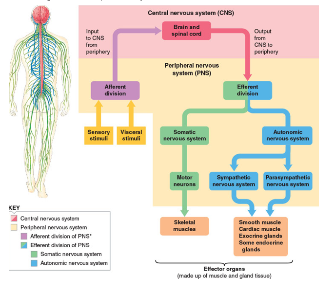
2. Organization of the nervous system
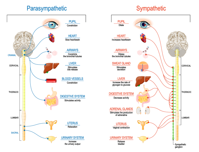
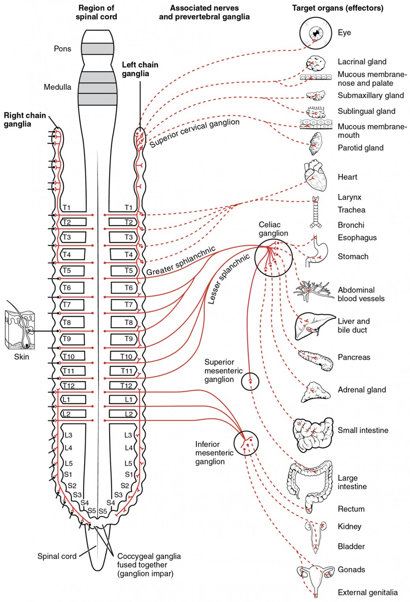
The autonomic nervous system exerts influence over the organ systems of the body to upregulate and downregulate various functions. The two aspects of the ANS operate as opposing functions that act to achieve homeostasis. The sympathetic nervous system, also known as the “fight or flight” system, increases energy expenditure and inhibits digestion. The following changes take place on activation of the sympathetic nervous system:
• Heart rate and cardiac muscle contractility increase
• The ciliary muscle relaxes, and the pupil becomes dilated for the betterment of far vision.
• Bronchodilation of the lungs
• Decreased GI motility
• Decreased urine output
• Relaxation of the detrusor muscle of the bladder and contraction of urethral sphincters
• Increased secretions from sweat glands
• Increased blood flow to muscles because of relaxation of arterioles
• Constriction of large arteries and large veins
• Increased metabolism, increased glucose production and mobilization by the liver
• Increased lipolysis within fat tissue
• Suppression of the immune system
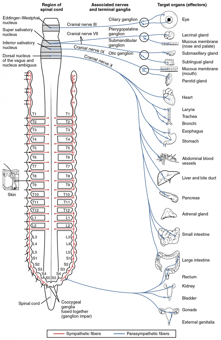
The parasympathetic nervous system, also known as “rest and digest,” can be thought of as functioning in opposition to the sympathetic nervous system. Its functions include:
• A decrease in heart rate and contractility of cardiac muscle
• Constriction of the ciliary muscle and the pupil for near vision
• Increased secretion by lacrimal glands and salivary glands
• Increased gut motility, bronchoconstriction of the lungs
• Contraction of the detrusor muscle with the relaxation of urethral sphincters
• Glycogen synthesis by the liver
• Swelling of the clitoris and erection of the penis
• Activation of the immune system
3. Autonomic Ganglia
Autonomic ganglia are aggregations of neurones that lie along peripheral nerve trunks where synapses are made by the axons of preganglionic neurones projecting from the brainstem and spinal cord.
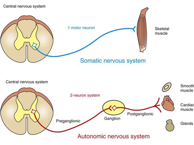
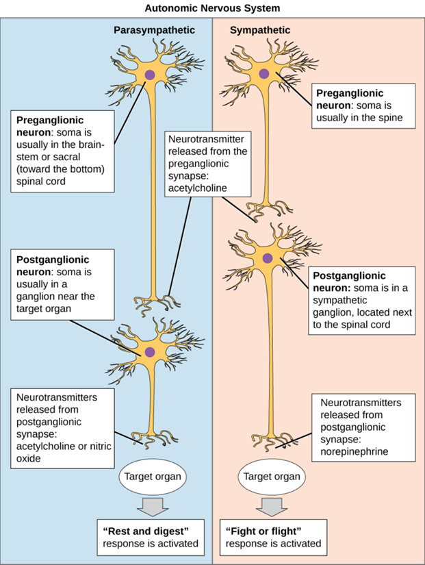
The sympathetic nervous system contains cell bodies that lie within the lateral gray column of the spinal cord running from T1 to L2. These neurons are known as preganglionic neurons and travel to ganglia, where they synapse and activate nicotinic receptors on postganglionic neurons using acetylcholine. The postganglionic neurons then travel to the target site and release norepinephrine to activate adrenergic receptors.
The parasympathetic nervous system is comprised of a cranial portion, consisting of cranial nerves III, VII, IX, and X and pelvic splanchnic nerves that exit from S2 to S4. The organization of the parasympathetic nerves is similar to the sympathetic nervous system, as preganglionic fibers synapse on postganglionic fibers, which then innervate target sites. A notable difference is that the preganglionic nerve fibers synapse on ganglia that are close to or embedded within their respective target sites, resulting in short postganglionic neurons.
4 the neurotransmitters of the autonomic ganglion
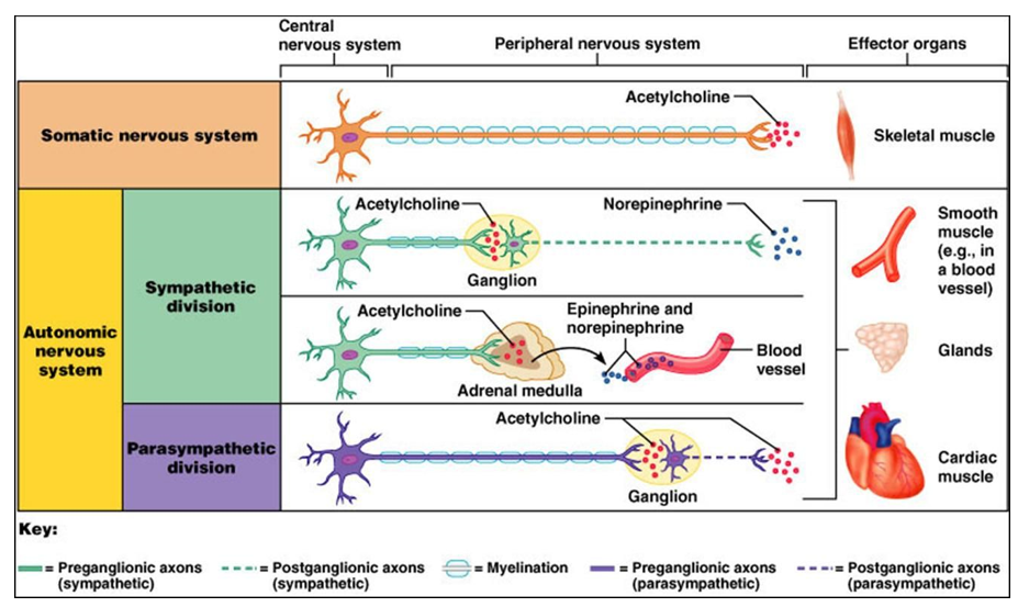
The neurotransmitters involved in the ANS are acetylcholine, norepinephrine, and epinephrine. Preganglionic neurons of the sympathetic and parasympathetic and postganglionic neurons of the parasympathetic nervous system utilize acetylcholine (ACh) (cholinergic synapses). Postganglionic neurons of the sympathetic nervous system use norepinephrine and epinephrine (adrenergic synapses)
5. Postsynaptic receptors of ANS
ACh is released by all preganglionic neurons, regardless of their involvement in the parasympathetic or sympathetic nervous system. ACh binds to nicotinic acetylcholine receptors, a ligand-gated ion channel, on postganglionic neurons. Once ACh binds, the ion channel is open to allow the flow of ions, specifically sodium and calcium, which induces an excitatory postsynaptic potential, and results in the propagation of a neuronal signal down the axon. ACh also gets released by postganglionic neurons of the parasympathetic nervous system. Here, ACh binds to muscarinic acetylcholine receptors on the target site, which are G-protein-coupled receptors. . It is important to note that ACh also gets released by postganglionic neurons of sweat glands and binds to and activates muscarinic receptors. This activity is an exception to the sympathetic nervous system, which normally involves norepinephrine as its chemical messenger released from postganglionic neurons
Norepinephrine gets released by postganglionic neurons of the sympathetic nervous system, which binds to and activates adrenergic receptors, which are G-protein-coupled receptors
Epinephrine is released into systemic circulation by chromaffin cells within the adrenal gland, specifically the adrenal medulla. The sympathetic nervous system splanchnic nerves synapse on chromaffin cells and stimulate them to release epinephrine using the signal messenger acetylcholine. This feature of the adrenal gland is an important component of the sympathetic nervous system as it allows systemic effects to occur on major organ systems
1.1 Acetylcholine receptors
There are two subtypes of nicotinic receptors, the muscular type (N1) and the neuronal type (N2).. The N1 receptor is present on skeletal muscle at the neuromuscular junction. N2 is within the peripheral and central nervous systems. N2 receptors are on the cell bodies of postganglionic neurons within the parasympathetic and sympathetic nervous systems. They are also on the adrenal medulla as a component of the sympathetic nervous system.
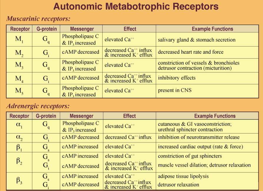
There are five different types of muscarinic receptors, M1, M2, M3, M4, and M5. Furthermore, each subtype is either stimulatory or inhibitory. Subtypes M1, M3, and M5 are stimulatory function through the phospholipase C second messenger pathway, while M2 and M4 function through a second messenger pathway that inhibits adenylate cyclase and prevents the formation of cAMP from ATP. The M1 muscarinic receptor is in the cerebral cortex, salivary, and gastric glands. M2 receptors are present in smooth muscle as well as cardiac tissue. M3 receptors are in smooth muscle cells, particularly of the bronchioles, iris, bladder, and small intestines. M4 and M5 receptors have a less clear distribution but have been found in the hippocampus, substantia nigra, and other locations within the brain
1.2 adrenergic receptors
The two major categories of adrenergic receptors (AR) are designated alpha and beta. Each has sub types: Receptor α1 involves a Gq protein that elevates C++, resulting in smooth muscle contraction. Receptor α2 involves a Gi protein that decreases cAMP and C++ influx which inhibits neurotransmission. Receptors β1, β2 & β3 involve a Gs protein and cAMP elevation that increases C++ for smooth muscle contraction (β2 & β3 can be linked to a Gi protein that decreases cAMP resulting in smooth muscle relaxation). Thus, the effect of ACh and NE can be either excitory or inhibitory, based on the receptor distribution within visceral organs.
6 The acetylcholine synaptic transmission
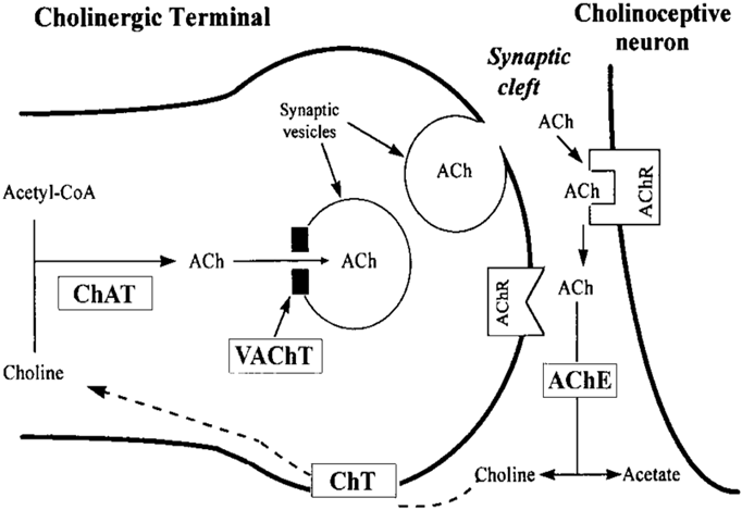
Synthesis and storage: Acetylcholine is synthesized from choline and acetyl coenzyme A by the enzyme choline acetyltransferase then stored in synaptic vesicles.
Release: An action potential reaching the nerve ending triggers an influx of calcium ions, provoking the release of ACh by exocytosis.
Binding to receptors: ACh diffuses across the synaptic cleft to bind with either nicotinic or muscarinic receptors on the postsynaptic neuron.
Breakdown and recycling: the enzyme acetylcholinesterase (AChE) hydrolyzes ACh into choline and acetate. Choline is then actively transported back into the presynaptic terminal; then recycled
7 The norepinephrine synaptic transmission
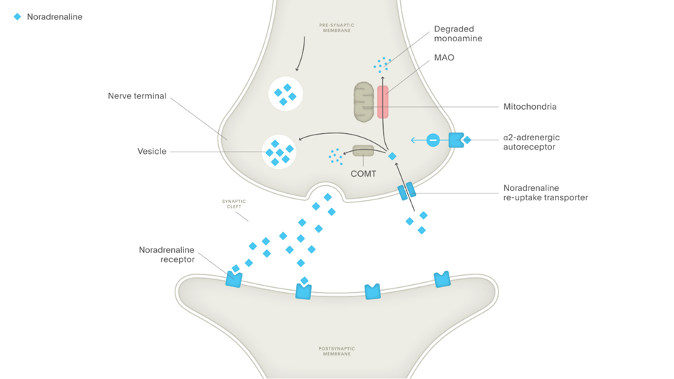
Synthesis and storage: Norepinephrine is synthesized from dopamine in the presynaptic neuron and transported by the vesicular monoamine transporter (VMAT).
Release: An action potential reaching the nerve ending triggers an influx of calcium ions, provoking the release of Norepinephrine by exocytosis.
Binding to receptors: Norepinephrine diffuses across the synaptic cleft to bind with specific adrenergic postsynaptic neuron
Breakdown and recycling: Norepinephrine reuptake by the norepinephrine transporter (NET), which pumps it back into the presynaptic neuron. Once inside, it can either be repackaged into vesicles by VMAT for future release or degraded by the enzymes monoamine oxidase (MAO) or catechol-O-methyltransferase (COMT).
8 functioning of the postganglionic fibers