Introduction:
Synapses are specialized areas of contact between neurons, between sensory cells and neurons, or between neurons and effector cells. Synapses mediate communication between cells
Synaptic transmission refers to the process in which a chemical signal is released from one neuron and diffuses to other neurons or target cells, generating a signal that excites, inhibits, or modulates cellular activity.
Classification
1-According to the nature of the connections:
Neuro-neuronal synapses: Contact points between two nerve elements,
Neuro-effector synapses: Consisting of a presynaptic element (axon terminal) and a postsynaptic element: smooth or striated muscle fiber or glandular cell
2- According to the postsynaptic effect: Excitatory synapses or Inhibitory synapses
3-According to the mode of function:
Electrical synapses: These allow electrical and metabolic coupling between cells through specialized contact zones called gap junctions. the synaptic cleft is very small (a few nanometers), transmission is very rapid and bidirectional
Chemical synapses: These require a chemical intermediary (neurotransmitter) between the pre- and postsynaptic elements. The synaptic cleft is wider, and transmission is unidirectional and significantly slower
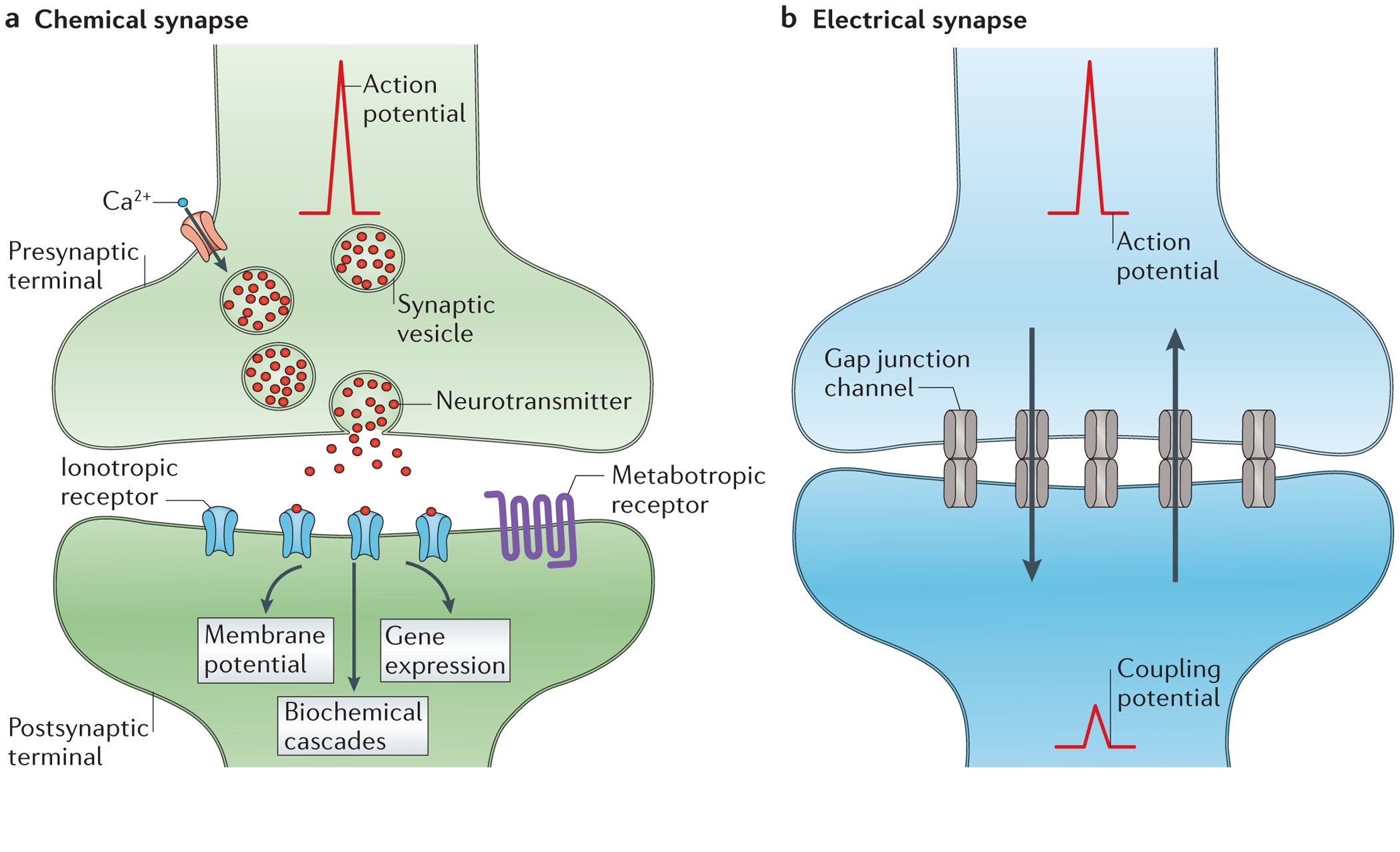
Synapse
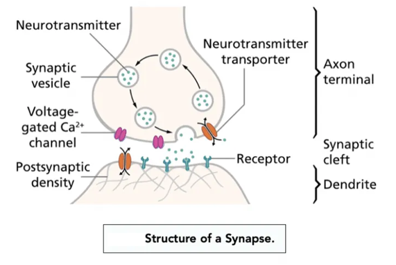
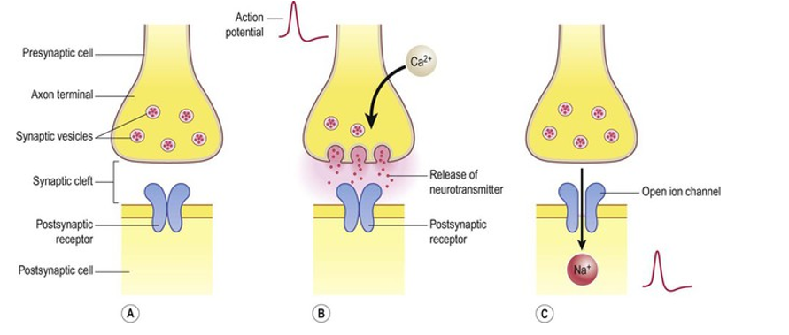
A synapse has a tripartite structure consisting of a presynaptic compartment, synaptic cleft, and postsynaptic compartment. The presynaptic terminal contains vesicles filled with neurotransmitters, which are released into the synaptic cleft, a tiny gap between the two neurons. The neurotransmitters then diffuse across the cleft to bind to receptors on the postsynaptic membrane, transmitting the signal
synaptic transmission at Neuromuscular Junction
Motor end plate, or neuromuscular junction, is defined as the specialized attachment site where a motor nerve fiber connects to the skeletal muscle fiber, characterized by unmyelinated axon terminals that release the neurotransmitter acetylcholine into synaptic clefts. The mechanism of neurotransmission at the neuromuscular junction is a finely tuned process that includes several steps, this process begins with action potential propagation, muscle contraction and acetylcholine breakdown
Action Potential Propagation to the Nerve Terminal: An action potential travels down the motor neuron and reaches the nerve terminal. The voltage change causes the voltage-gated calcium channels in the nerve terminal membrane to open.
Calcium Influx and Vesicle Fusion: Calcium enters the nerve terminal and binds to synaptotagmin, causing a conformational change in the latter that allows it to interact with SNARE proteins. This interaction triggers the fusion of synaptic vesicles with the presynaptic membrane, releasing ACh into the synaptic cleft via exocytosis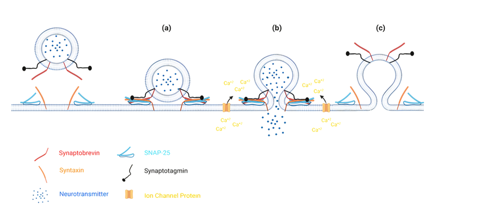
Acetylcholine Binding to Receptors: ACh then diffuses across the synaptic cleft and binds to the nAChRs located on the motor end plate of the muscle fiber. ACh binding opens the receptor channels, allowing sodium ions to enter the muscle cell, thus depolarizing the membrane.
Generation of Endplate Potential: The influx of sodium ions depolarizes the postsynaptic membrane, shifting the potential from -90 mV to -40 mV and creating a local depolarization known as the endplate potential (EPP). A single EPP is sufficient to reach the threshold and generate an action potential that propagates along the muscle fiber
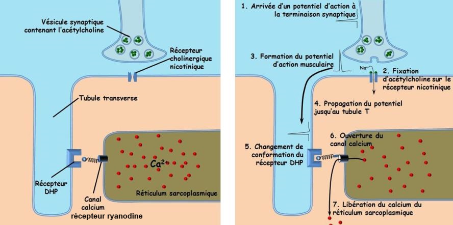
Muscle Contraction: The action potential travels deep into the muscle fiber through the transverse tubules (T-tubules), which are invaginations of the sarcolemma. The dihydropyridine (DHP) receptor, located in the T-tubule membrane, detects the depolarization of the sarcolemma, producing a conformational change in the DHP receptor.The DHP receptor is mechanically coupled to the ryanodine receptor located on the sarcoplasmic reticulum membrane. The conformational change in the DHP receptor triggers the opening of the ryanodine receptor, allowing calcium ions to be released from the sarcoplasmic reticulum into the muscle cell's cytoplasm. The released calcium causes muscle contraction.
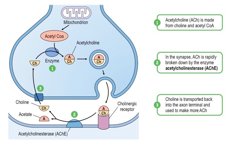
Acetylcholine Breakdown: ACh is rapidly broken down by acetylcholinesterase (AChE) in the synaptic cleft after binding to nAChRs and triggering muscle depolarization, preventing continuous stimulation of the muscle fiber. The breakdown products are then taken up into the presynaptic terminal for recycling
Neuro-neuronal synapses
A neuro-neuronal synapse is a point of communication where the axon terminal of a presynaptic neuron communicates with a postsynaptic neuron (dendrite, soma, or axon) including axodendritic synapses, axo-somatic synapses, axo-axonic synapses
Synaptic transmission is the process at synapses by which a chemical signal (a transmitter) is released from one neuron and diffuses to other neurons where it generates a signal which excites, inhibits or modulates cellular activity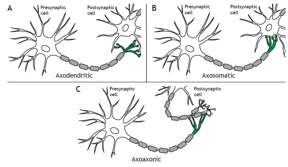
Excitatory and inhibitory postsynaptic potential
An excitatory postsynaptic potential (EPSP) is the change in membrane voltage of a postsynaptic cell following the influx of positively charged ions into a cell (typically Na+) because of the activation of ligand-sensitive channels. This results in a depolarization of the postsynaptic cell, thus increasing the likelihood of action potential propagation. These excitatory synapses serve to increase excitability in neurones. Conversely, inhibitory postsynaptic potentials (IPSP) result from the influx of negative ions (e.g., Cl−) into, or the efflux of positive ions (e.g., K+), out of the postsynaptic cell. This results in cell hyperpolarization and thus decreases the likelihood of action potential propagation, and therefore represent inhibitory synapses.
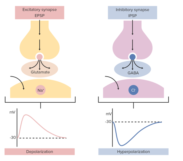
Electrophysiological characteristics of EPSP and IPSP
Depolarization or Hyperpolarization of postsynaptic membrane.
Localized: EPSPs and IPSPs occur at the postsynaptic membrane, usually on dendrites or the soma (the depolarization or hyperpolarization is not generated everywhere, only in the small region of membrane where the ligand-gated channels open)
Passive decay: EPSPs and IPSPs are electrotonic potentials that decay over both time and distance. The further a synapse is from the cell body, the smaller the EPSP/ IPSPs will be at the axon hillock
Duration : depends on the type of receptor for example: AMPA receptors (Ionotropic) for glutamate lasts only a few milliseconds which produce fast synaptic transmission. However metabotropic glutamate receptors, open channels indirectly last hundreds of milliseconds to seconds which produce slow synaptic transmission.
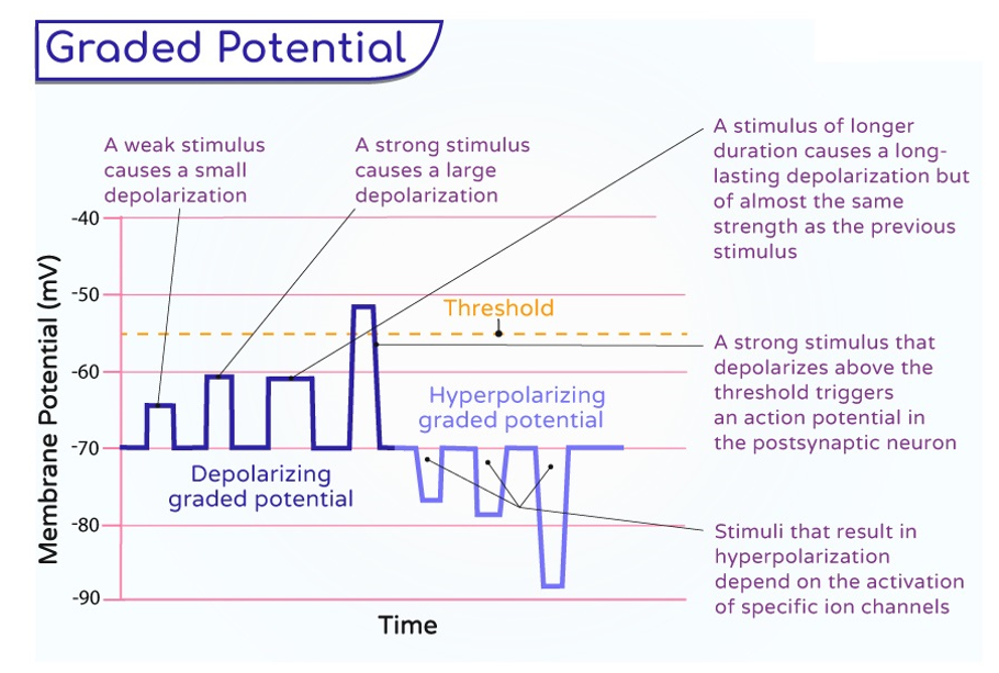
Graded potentials : are changes in the electrical charge across a neuron's membrane that vary in size, rather than being all-or-none events (for EPSP), depends on the amount of neurotransmitter released and the number of channels that open.
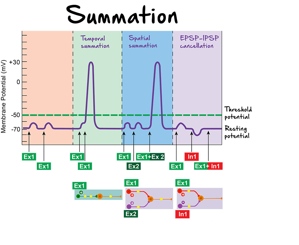
Summation : the process by which a postsynaptic neuron integrates several EPSPs in a temporal or spatial manner to reach the threshold of an action potential. The EPSP signal is propagated down the dendrite and is summed with other inputs at the axon hilllock.
Temporal Summation: occurs when a single synapse releases neurotransmitter several times in rapid succession. Each EPSP adds to the next before the previous one has fully dissipated. If these successive depolarizations accumulate sufficiently, they can bring the membrane potential to threshold and trigger an action potential.
Spatial summation: occurs when stimuli are applied at the same time, but in different areas (different synapses on the same neuron) , with a cumulative effect upon membrane potential.
Cancellation : occurs when EPSP and IPSP reach the postsynaptic neuron at the same time. EPSPs depolarize the membrane, while IPSPs hyperpolarize it. As a result, an IPSP can partially or completely cancel the depolarizing effect of an EPSP, preventing the membrane from reaching the threshold and stopping the generation of an action potential.
Neurotransmitters
Neurotransmitters are endogenous chemicals that allow neurons to communicate with each other throughout the body. They enable the brain to provide a variety of functions, through the process of chemical synaptic transmission. These endogenous chemicals are integral in shaping everyday life and functions.
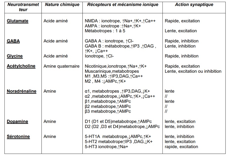
Receptor Activation: Once the neurotransmitter binds to the post-synaptic neuron, it can generally activate 1 of 2 types of receptors: a ligand-gated ion channel or a G-protein receptor.
Ligand-Gated Ion Channel: When the neurotransmitter binds to this receptor, the attached ion channel has a direct opening or closing. In other words, the neurotransmitter acts directly on the target ion channel. This receptor type is described as “fast” because it generally only takes a few milliseconds to produce a response and is terminated very quickly. These receptors can be excitatory or inhibitory, depending on which neurotransmitter is binding to the receptor.
G-Protein Coupled Receptors: These receptors produce a response (opening or closing an ion channel) by activating a signaling cascade involving secondary messengers. The most common secondary messengers are cyclic adenosine monophosphate (cAMP), inositol triphosphate (IP3), and diacylglycerol (DAG). When the neurotransmitter binds to the receptor, it activates the G-protein, which binds to guanosine triphosphate (GTP) and is activated. This activates the secondary messenger cascade, eventually leading to the phosphorylation of ion channels. Due to multiple steps having to take place to generate the final response, this pathway is generally described as “slow,” and the effects last longer (seconds to minutes).
There are a number of neurotransmitters used by the body for different functions, including:
Acetylcholine (ACh): One of the most important neurotransmitters found in multiple synapses in the body, including, the neuromuscular junction, autonomic ganglia, caudate nucleus, and the limbic system. Generally, ACh is an excitatory neurotransmitter at the neuromuscular junction and in the autonomic ganglia.
Glutamate : is the principal excitatory neurotransmitter used in the brain, essential for the transmission of nerve impulses between neurons, particularly in learning and memory.
Gamma-aminobutyric acid (GABA) and glycine, conversely, serve as the major inhibitory neurotransmitters. GABA, for example, can account for approximately 40% of the inhibitory processing in the brain. Glycine is found primarily in the spinal cord
Dopamine, another major neurotransmitter, plays an essential role in several brain functions, including learning, motor control, reward, emotion, and executive functions. Dopamine has also been implicated in psychiatric and neurological disorders. Dopamine is both an excitatory and inhibitory neurotransmitter. It can excite neurons, for example by opening sodium channels, and inhibit them, notably by acting on receptors that open potassium channels. Its action depends on the type of dopaminergic receptor involved and the region of the brain.
Serotonin is a neurotransmitter which can be both excitatory and inhibitory, its effect depending on the type of receptor to which it binds, that modulates multiple neuropsychological processes and neural activity many drugs used in psychiatry and neurology target serotonin. Serotonin also has implications that affect gastrointestinal processes, bladder control, and cardiovascular function.
Noradrenaline is primarily an excitatory neurotransmitter, in the brain exerts effects on a variety of processes, including stress, sleep, attention, focus, and inflammation. It also plays a role in modulating the responses of the autonomic nervous system.
Adrenaline is an excitatory neurotransmitter that prepares the body for a fight response by increasing heart rate, blood pressure, and alertness.
Endorphins are primarily inhibitory neurotransmitters because they have an analgesic effect, meaning they block the transmission of pain signals