
Osmolarity and Plasma Osmolarity
The relative proportions of solutes and solvents are expressed in: Grams/l , Moles/l, Equivalents/l, Osmoles/l (or /kg of solvent).
Osmolarity is the measure of the total concentration of dissolved particles in a solution. It represents the number of osmoles of solute per liter of solution (Osm/L).
Osmolarity of one solution relative to another
– Iso-osmotic: same amount of solute per volume
– Hyperosmotic: higher
– Hypo-osmotic: lower
Plasma osmolarity is a measurement of the total concentration of dissolved solutes in the blood plasma, primarily sodium, glucose, and urea. It represents the plasma’s ability to attract water through osmosis.
Plasma osmolarity depends mainly on serum sodium, which accounts for almost half of the total osmoles in plasma (about 140 mmol/L).Therefore, to quickly estimate plasma osmolarity, you can simply double the serum sodium concentration, giving an approximate value of 280 mOsm/L.
If we take into account non-electrolytes
Plasma osmolality = 2 × [Na+] + [glucose] + [urea]
plasma osmolality formula is 2[Na+] + [Glucose]/18 + [urea]/2.8 (if Concentrations of glucose and urea are expressed in mg/dL)
Normal plasma osmolarity: around 285–295 mOsm/L
Tonicity
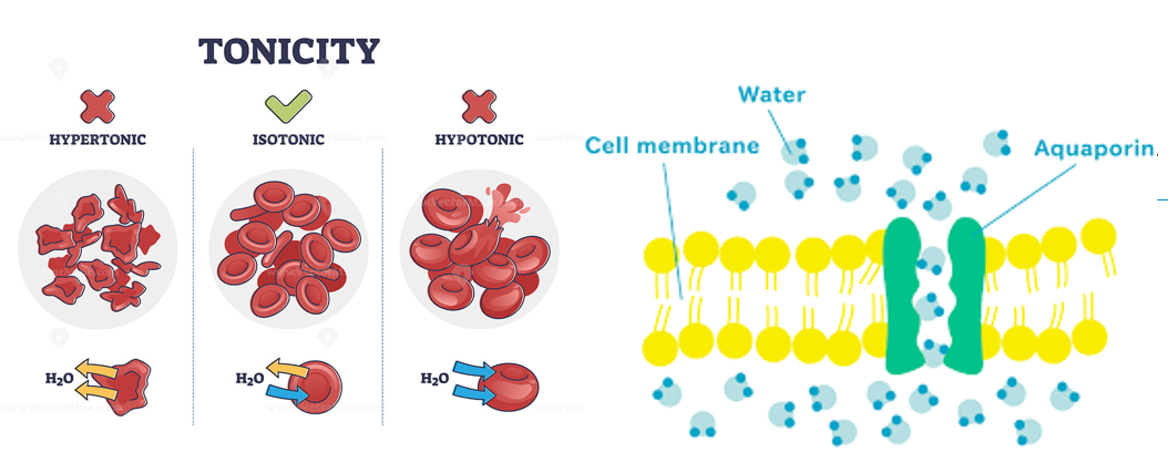
Tonicity refers to the effect of a solution’s osmolarity on cell volume; that is, how a solution influences the movement of water across a cell membrane.
A hypotonic solution causes cells to swell (water enters the cell).
An isotonic solution causes no change in cell volume.
A hypertonic solution causes cells to shrink (water leaves the cell).
Exchanges between fluid compartments
Exchanges between the plasma and the external environment
Humans absorb and eliminate water, sodium, potassium, and phosphorus through various exchange organs. Under normal conditions, the net effect of these exchanges is zero, meaning that the overall balance of these substances is maintained.
Plasma circulates rapidly throughout the body and is in continuous contact with the external environment via the skin, digestive tract, respiratory system, and kidneys.
There
are numerous normal variations in water and electrolyte balance
depending on the individual
and specific circumstances.
However, urinary excretion is constantly regulated to maintain overall water and electrolyte equilibrium.
Exchanges between plasma and interstitial fluid
Capillaries are semi-permeable structures providing a surface area of about 300 to 1000 m² for exchange between the plasma and the interstitial fluid.
Exchange by diffusion:
This process is continuous,
occurring at approximately 60 L/min,
and is bidirectional. It allows the rapid and balanced exchange of blood gases
and small molecules between plasma and interstitial fluid.
Exchange by filtration and reabsorption:
This is the main mechanism of fluid exchange, as it can vary dynamically and thereby modify the volumes of both compartments.
It is governed by the balance of: Capillary and interstitial
hydrostatic pressures, and Capillary and interstitial oncotic
pressures.
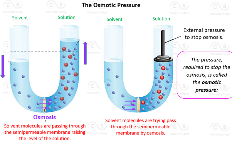
Osmotic pressure is the minimum pressure needed to prevent the net the movement of water across a semipermeable membrane
Oncotic pressure is the part of the osmotic pressure in the blood plasma that is generated by plasma proteins, mainly albumin. These proteins are too large to cross capillary walls, so they attract water and help keep it inside the blood vessels. This process is known as reabsorption
Hydrostatic Pressure
Hydrostatic pressure is the pressure that blood or interstitial fluid exerts on the capillary walls.
Capillary hydrostatic pressure (Pc):
The pressure exerted by the blood inside the capillaries.
It pushes water out of the capillaries into the interstitial
space.
Interstitial hydrostatic pressure (Pi):
The pressure in the interstitial fluid
surrounding the capillaries. Pushes fluid into the capillary
Capillary hydrostatic pressure is typically much higher than interstitial hydrostatic pressure,
which causes a net movement of fluid from the
capillaries into the tissues.
This process is known as filtration
In the first half of the capillary (arterial pole):
Hydrostatic pressure > oncotic pressure,
causing water and dissolved substances to move from the plasma into the interstitial fluid (filtration).
In the second half of the capillary (venous pole):
Oncotic pressure > hydrostatic pressure,
causing water and solutes to return to the plasma (reabsorption).
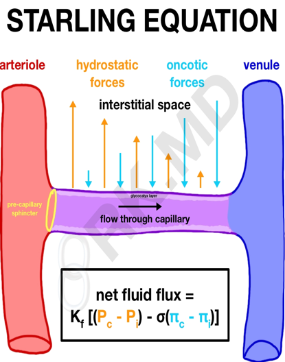
Kf :filtration coefficient
Pc :capillary hydrostatic pressures
Pi :interstitial hydrostatic pressures
σ : reflection coefficient
πc :capillary oncotic pressures
πi :interstitial oncotic pressures
Kf :filtration coefficient
Pc :capillary hydrostatic pressures
Pi :interstitial hydrostatic pressures
σ : reflection coefficient
πc :capillary oncotic pressures
πi :interstitial oncotic pressures
Exchanges between fluid compartments
Exchanges between the intracellular and extracellular compartments
Water exchanges
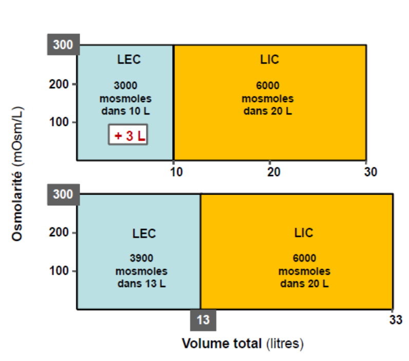
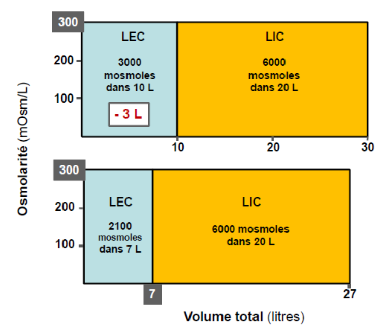
Under normal conditions, osmolarity is nearly identical in all body compartments. However, the intracellular fluid volume is approximately twice that of the extracellular fluid volume.
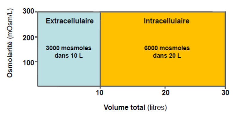
If
there is a gain
(e.g., isotonic saline infusion) or a loss of isotonic fluid
(e.g., severe diarrhea), there will be no movement of water
between the intracellular and extracellular compartments and no change in osmolarity.
However, there will be a change in extracellular fluid
volume,
it will increase with isotonic gain and decrease with isotonic loss.
In
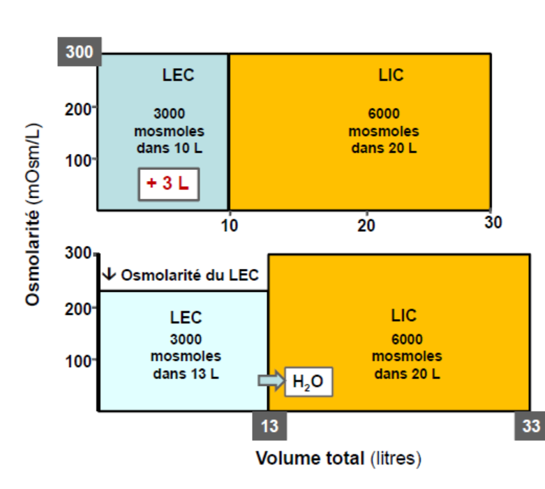
the case of a gain of pure water,
there will first be an increase in extracellular volume
and a decrease in extracellular osmolarity.
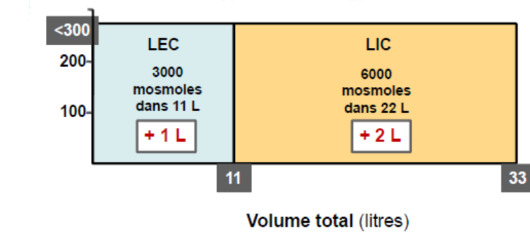
This osmotic difference causes water to move from
the extracellular compartment into the intracellular compartment until a new equilibrium is reached.
At this point, both intracellular and extracellular volumes increase, while osmolarity decreases
in both compartments.
In
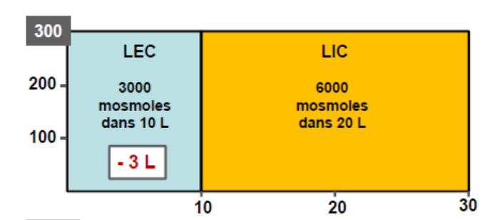
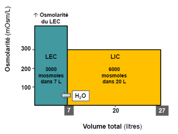
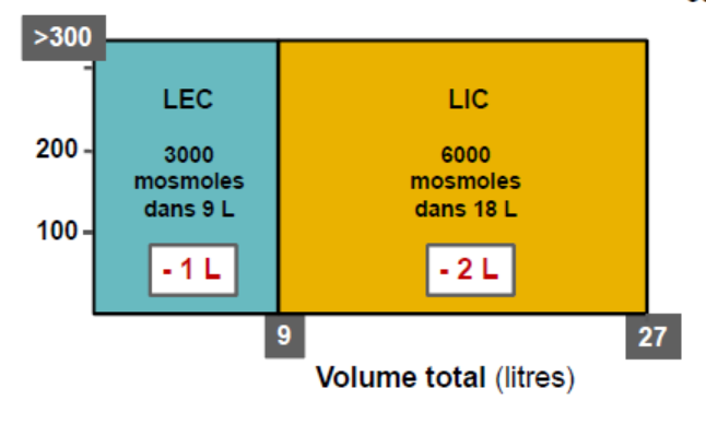
the case of a loss of pure water
(as in diabetes insipidus), there will be a decrease in extracellular volume
and an increase in extracellular osmolarity.
This osmotic imbalance causes water to move from
the intracellular compartment into the extracellular compartment to restore equilibrium.
As a result, both intracellular and extracellular
volumes decrease, while osmolarity increases
in both compartments.
Electrolyte movement
Passive Transport by Diffusion
Ions move down their chemical or electrical gradients without energy expenditure.
Simple diffusion: occurs directly across the plasma membrane for blood gases (O₂, CO₂) and some small lipid-soluble molecules.
Ion diffusion: takes place through selective ion channels that allow specific ions (e.g., Na⁺, K⁺, Cl⁻) to pass.
Facilitated diffusion: uses membrane transporters (carriers or permeases) to move larger or polar molecules such as glucose down their concentration gradient.
Active Transport
Active transport moves substances against their concentration or electrical gradients and requires energy.
Primary active transport: uses ATP directly, as in the Na⁺/K⁺-ATPase pump, which maintains the ionic difference between the intracellular and extracellular environments.
Secondary active transport: uses the energy stored in ionic gradients to drive the co-transport of other substances (e.g., glucose–Na⁺ cotransport).
Metabolic acidosis is caused by either an overproduction of acid (H⁺) in the body for example, in diabetic ketoacidosis or by a loss of bicarbonate (HCO₃⁻) through severe diarrhea or kidney failure. As a result, [H⁺] increases in the extracellular fluid , and the blood becomes more acidic.to protect the blood pH, H⁺ ions move into the cells. Because cells must remain electrically neutral, K⁺ ions move out of the cells into the blood. This leads to hyperkalemia (high serum potassium), which can cause cardiac arrhythmias.
Metabolic alkalosis occurs due to either a loss of acid (H⁺) such as with prolonged vomiting or diuretic use or an excess intake of bicarbonate, for example, from overconsumption of antacids or bicarbonate-containing drugs. In this case, [H⁺] decreases in the ECF, making the blood more alkaline. To maintain balance, H⁺ ions move out of the cells into the blood. To preserve electrical neutrality, K⁺ ions move into the cells. This results in hypokalemia (low serum potassium), leading to muscle cramps, weakness, and cardiac arrhythmiaswater imbalances
Dehydration (Increased ECF Osmolarity :Hypertonicity)
Causes: Dehydration can result from, diabetes insipidus, lack of water intake…, cause excessive loss of water and electrolytes from the body.
Physiological Effects: Water loss occurs first from the
extracellular fluid (ECF).
As the ECF becomes hyperosmotic, water moves out of the cells to restore
osmotic balance so cell shrinkage. This leads to a decrease in both
extracellular and intracellular volumes, affecting cell function and
potentially leading to circulatory collapse.
hyperhydration: (Decreased ECF Osmolarity: Hypotonicity)
Causes: kidney failure or excessive absorption of a large quantity of water….etc
Signs: hyponatremia (too low a concentration of sodium in the blood). This excessive dilution of sodium in the interstitial fluid will, through osmosis, cause water to enter the cells, which will swell. It is urgent to inject a hypertonic saline solution (highly concentrated) to draw water from the cells and thus reverse the osmotic gradient, otherwise nausea, vomiting, muscle cramps, cerebral edema…etc
EDEMA
Edema is the abnormal accumulation of fluid in the interstitial spaces. It occurs when filtration exceeds reabsorption across capillary walls, disturbing the balance between hydrostatic and oncotic pressures.
Most plasma proteins, especially albumin, are synthesized in the liver. In alcoholic cirrhosis or severe liver disease, protein synthesis decreases, leading to a reduction in plasma oncotic pressure. As a result, more fluid leaves the capillaries than is reabsorbed, causing water accumulation in the interstitial space, and hence edema.
2. Edema due to decreased plasma oncotic pressure (malnutrition)
In cases of severe malnutrition (e.g., kwashiorkor), the diet lacks essential amino acids required for plasma protein synthesis. This leads to hypoproteinemia (low serum protein), decreased oncotic pressure, and generalized edema.
3. Edema due to kidney damage
In nephrotic syndrome or glomerular damage, the kidneys lose large amounts of albumin in the urine (proteinuria). Although protein synthesis is normal, the loss of plasma proteins causes a drop in oncotic pressure, leading to edema, often first visible in the face or around the eyes.
4. Edema due to increased capillary hydrostatic pressure
In heart failure, leading to increased hydrostatic pressure in capillaries, especially on the venous side. This promotes excess filtration of fluid into tissues. In right heart failure, edema mainly affects the lower limbs. In left heart failure, blood backs up in the lungs, causing pulmonary edema
5. Edema due to lymphatic obstruction
Normally, the lymphatic system drains excess interstitial fluid. If the lymphatic vessels are obstructed, fluid accumulates locally, causing lymphedema. A classic example is elephantiasis, common in intertropical regions, caused by parasitic filarial worms transmitted by mosquito bites. The lymphatic blockage leads to massive swelling of affected limbs or genitals.