
1 Extraction and purification of nucleic acids
Any study of molecular genetics involves the provision of a sample of nucleic acids. The techniques of extraction of nucleic acids are relatively simple. It should be simply to avoidany destruction enzymatic or mechanical. In fact, the nucleic acids that are stable in the intact cell, become very vulnerable to digestion by nucleases endogenous once the cell lysée.
The protocol for extraction of genomic DNA from a bacterial culture is based on the principle of alkaline lysis. After extraction, the DNA is resuspendu in 30 µl of TE and stored at -20°C. genomic DNA is analyzed by agarose gel electrophoresis in 1% (w/v). The protocol for DNA extraction is the following :
1. Transfer 1 ml of a préculture overnight at 37°C in an eppendorf tube
2. Centrifuge for 1 min at 13000 rpm
3. Remove the supernatant
4. Resuspend the pellet in 500 µl of a solution of TE (10 mM Tris-HCl pH 8.1 mM
EDTA, pH 8)
5. Add 20 µl lysozyme (50 mg / ml) and incubate 30min at 37 °C
6. Add 60 µl of EDTA (0.5 M, pH 8)
7. Add 10 µl Proteinase K (20mg/ ml) and incuber1 h at 37°C
8. Add 100 µl SDS 10% and incubate for 10 min at 50°C
9. Add 350 µl of acetate of potassium 3M pH 5.2
10. Mix gently by inversion of the tube, incubate for 10 min in ice
11. Centrifuge for 10 min at 10000rpm
12. Transfer the surnagent in an eppendorf tube nine
13. Add 1 volume of isopropanol
14. Mix by inversion (up to the appearance of the DNA in the form of a filament) and
leave to precipitate for 10 min at – 20 °C
15. Centrifuge for 1 min at 10000 rpm
16. Remove the supernatant
17. Add 200 µl of ethanol 70% to wash the DNA, no agitation
18. Centrifuge for 10 min at 10,000 rpm
19. Remove the supernatant
20. Dry the pellet by inverting the tube on a filter paper for 10 min
21. Dissolve the pellet in 20 µl TE.
6.2 determination of nucleic acids
The pyrimidines and the purines strongly absorb the UV light at 260nm.A unit of optical density at 260 nm corresponds to :
- A solution of DNA double-strand 50µg/ml.
- A solution of DNA single-strand or RNA to 25
C = A260 * DF * 100
These values apply to nucleic acids, perfectly pure and homogeneous solution.
To verify the purity of the DNA it is necessary to calculate : P (purity)= A260 /A280 ratio of A solution of DNA is considered pure if : 1.7 ≤ P ≤ 2
2 Electrophoresis
Although the sequence of the nucleic acid determines its biological functions, the length of these polymers is their physical property to be the most useful.Among the most commonly used techniques for the separation of nucleic acids according to their size is electrophoresis.
It is a technique of bioseparations for the separation of biomolecules according to their charge and their size (DNA, RNA, proteins). The term electrophoresis comes from : électroce which means electric energy and phoresis (photo in Greek) which means to carry, to have self-esteem.
The nucleic acids in solution are negatively charged due to the ionization of their groupings phosphate (macromolecules polyanionic evenly loaded). They also move towards the electrode (+). The sequences of nucleic acids have a ratio charge/mass almost identical, regardless of their length, because all the nucleotides are nearly the same mass (molar weight average molecular nucleotides 330 g/mol).
According to the theories of the electrophoretic mobility μ in an electric field within
a gel gifted to be able to filter is:
Log μ= Logµo - Kr. C
Log µo : The mobility of the molecule in the liquid medium
C: Concentration of the gel
Kr: Coefficient of retardation due to the gel itself is a function of the molecular mass of the molecule).
Two kinds of matrix gel are used : agarose and polyacrylamide. The gel acts as a molecular sieve through which the DNA molecules in motion must pass ; the large molecules have more difficulty to pass through the mesh (pore) that are created by the network of microfibers and gel will migrate more slowly than smaller molecules. The speed of migration of the DNA is influenced by two parameters :
- Molecular mass (number of pb)
- The gel concentration
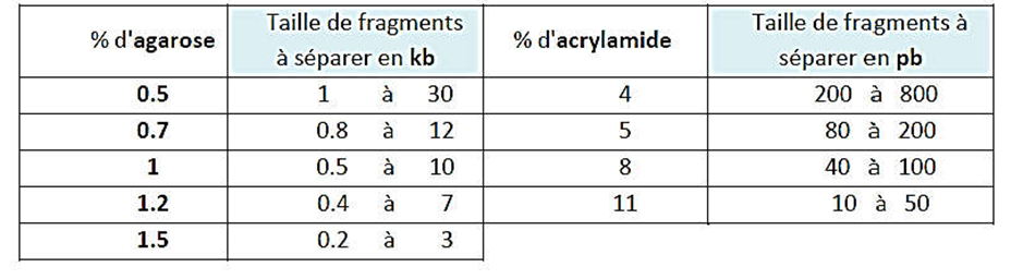
It should be noted that the nature and concentration of the media in electrophoresis are based on the size of the fragments to be separated.
Table 2: Size of the fragments separate according to the concentration of the gel.
In all electrophoresis, markers size (or molecular weight) are deposited and migrate along the DNA studied. Once the electrophoresis is complete, the molecules, fluorophores, such as ethidium bromide, which binds to DNA by intercalating between the bases. The bungs DNA (include all the DNA molecules of identical size) are visualized under UV.
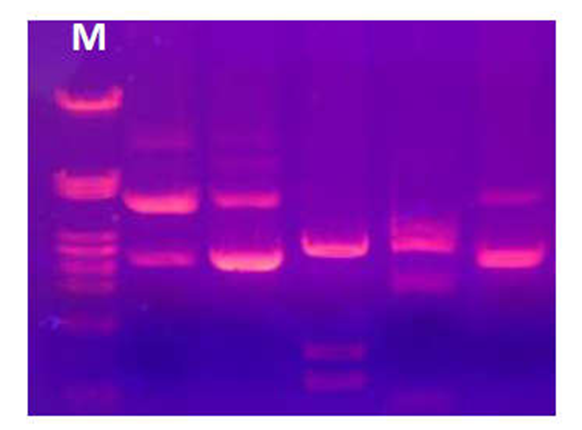
Figure 19: Visualization of bands of DNA under UV. M: marker size
The mobility relative is the ratio between the distance of migration of a band and the distance of migration of the front of migration.The right log(number of kb) = f(mobility), you can determine the size of a DNA molecule is unknown.