
1 The effects in which SANS acts in direct contrast to the PANS function
Eye
In the eye, sympathetic activation causes the radial muscle of the iris (alpha-1) to contract, which leads to mydriasis, allowing more light to enter. Furthermore, the ciliary muscle (beta-2) relaxes, allowing for far vision to improve.In the eye, parasympathetic stimulation of M3 receptors causes contraction of the iris's sphincter muscle, leading to pupil constriction (miosis). Additionally, it causes contraction of the ciliary muscle, improving near vision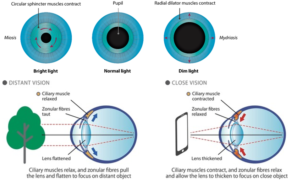
Heart
In the heart (beta-1, beta-2), sympathetic activation causes an increased heart rate, the force of contraction, and rate of conduction, allowing for increased cardiac output to supply the body with oxygenated blood.In the heart, parasympathetic stimulation of M2 receptors causes decreased heart rate and conduction velocity.
Lungs
In the lungs, bronchodilation (beta-2) and decreased pulmonary secretions (alpha-1, beta-2) occur to allow more airflow through the lungs. In the lungs, parasympathetic stimulation of M3 receptors leads to bronchoconstriction. It also increases bronchial secretions.
stomach and intestines
In the stomach and intestines, decreased motility (alpha-1, beta-2) and sphincter contraction (alpha-1), occur to slow down digestion to divert energy to other parts of the body.In the stomach and intestines, parasympathetic stimulation of M3 receptors leads to increased motility and relaxation of sphincters. Stimulation of M1 receptors also increases gastric secretions to aid in digestion.
Pancreas
The exocrine and endocrine pancreas (alpha-1, alpha-2) decreases both enzyme and insulin secretion.In the pancreas, parasympathetic stimulation of M3 receptors leads to the release of digestive enzymes and insulin.
Bladder
In the bladder, there is relaxation of the detrusor muscle and contraction of the urethral sphincter (beta-2-alpha1) to help stop urine output during sympathetic activation. In the bladder, parasympathetic stimulation of M3 receptors stimulates ureter peristalsis, contraction of the detrusor muscle, and relaxation of the internal urethral sphincter, aiding in the flow and excretion of urine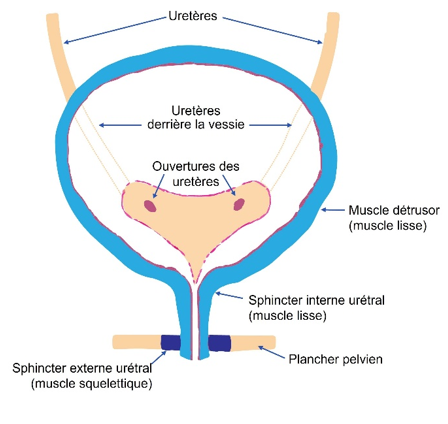
salivary glands
The salivary glands (alpha-1, beta-2) work through small volumes of potassium and water secretion In salivary glands, parasympathetic stimulation of M1 and M3 receptors leads to high-volume secretion of potassium ions, water, and amylase
2 Actions of SANS which do not oppose those of the PANS
Sympathetic function
• There is strong constriction through the alpha-1 receptor in arterioles of the skin, abdominal viscera, and kidney, and weak constriction through the alpha-1 and beta-2 receptors in the skeletal muscle.
• In the liver, increased glycogenolysis and gluconeogenesis (alpha-1, beta-2) occur to allow for glucose to be available for energy throughout the body.
• In the spleen, there is a contraction (alpha-1).
• Sweat glands(M3) and arrector pili muscles (alpha-1) work to increase sweating and erection of hair to help cool down the body.
• the adrenal medulla (nicotinic receptor) increases the release of epinephrine and norepinephrine to act elsewhere in the body
• The kidney (beta-1) increases renin secretion to increase intravascular volume.
Parasympathetic function
• In the male reproductive tract, parasympathetic stimulation of M3 receptors causes smooth muscle relaxation in the helicine arteries of the penis, allowing blood to fill the corpora cavernosa and corpus spongiosum, causing an erection. The PNS also gives excitatory signals to the vas deferens, seminal vesicles, and prostate.
• In the gallbladder, parasympathetic stimulation of M3 receptors stimulates contraction to release bile.
3 Parasympathetic nervous system control and heart function
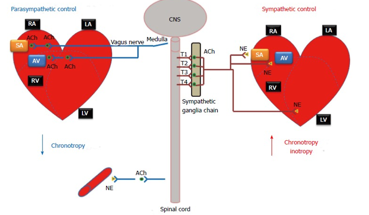
During rest, sleep, or emotional tranquility, the parasympathetic nervous system predominates and controls the heart rate at a resting rate of 60-75 bpm.
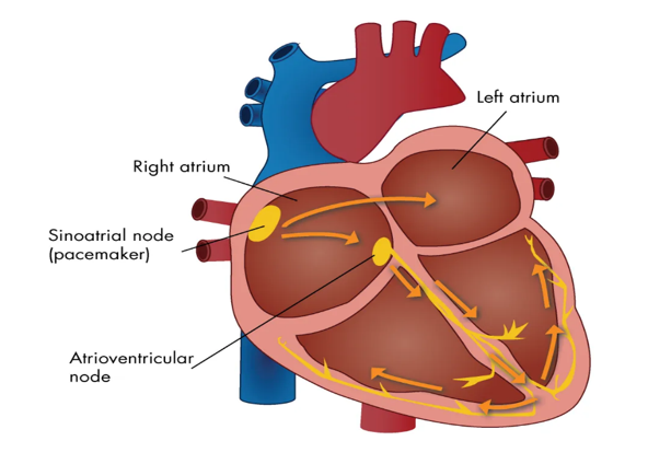
The M2 receptors are expressed in the heart; abundant in nodal and atrial tissue (The vagus nerve directly innervates the sinoatrial node), but sparse in the ventricles. The binding of acetylcholine to M2 receptors serves to slow heart rate till it reaches normal sinus rhythm. This is achieved by slowing the rate of depolarization (which increases the potassium current), as well as by reducing the conduction velocity through the atrioventricular node. Additionally, the activation of M2 receptors reduces the contractility of atrial cardiomyocytes, thus reducing, in part, the overall cardiac output of the heart as a result of reduced atrial kick, smaller stroke volume, and slower heart rate.
Negative chronotropic effect (decrease in heart rate)
Negative inotropic effect (decrease in myocardial contractility)
Negative dromotropic effect (decrease conduction velocity)
4 Sympathetic nervous system control and heart function
The sinoatrial (SA) node is the predominate pacemaker of the heart. It is located within the upper posterior wall of the right atrium and is responsible for maintaining a sinus rhythm of between 60 and 100 beats per minute. Myocardial contractility represents the ability of the heart to produce force during contraction. It is determined by the incremental degrees of binding between myosin (thick) and actin (thin) filaments, which in turn depends on the concentration of calcium ions (Ca2+) in the cytosol of the cardiomyocyte. Stimulation by the sympathetic nervous system causes an elevation in intracellular (Ca2+) and thus an increase in contraction of both the atria and ventricles.
Positive chronotropic effect (increase in heart rate)
Positive inotropic effect (increase of contractility)
Positive dromotropic effect (increase of conduction)
5 Adrenal medulla
The adrenal medulla is GIGANTIC. It’s the largest sympathetic ganglia. The outer layer is adrenal cortex which is an endocrine organ, while the inner layer is the medulla. When stimulated by preganglionic sympathetic fibers from T8-L1, they secrete large quantities of the excitatory hormones norepinephrine and epinephrine (adrenaline) into nearby capillaries. When these two hormones are released in the blood they amplify all of this fight or flight stuff to give you more energy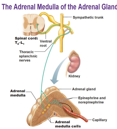
A mass sympathetic discharge, which typically occurs during the “fight-or-flight” response and during exercise, involves the simultaneous stimulation of organs and tissues throughout the body. Included among these tissues are the adrenal medullae which release epinephrine and norepinephrine into the blood. In large part, the indirect effects of these catecholamines are similar to and, therefore, reinforce those of direct sympathetic stimulation. However, there are some important differences in the effects of the circulating catecholamines and those of norepinephrine released from sympathetic nerves.
The duration of activity of the catecholamines is significantly longer than that of neuronally released norepinephrine. Therefore, the effects on the tissues are more prolonged. This difference has to do with the mechanism of inactivation of these substances. Norepinephrine is immediately removed from the neuroeffector synapse by way of reuptake into the postganglionic neuron. This rapid removal limits the duration of the effect of this neurotransmitter. In contrast, there are no enzymes in the blood to degrade the catecholamines. Instead, the catecholamines are inactivated by COMT in the liver. As one might expect, the hepatic clearance of these hormones from the blood would require several passes through the circulation. Therefore, the catecholamines are available to cause their effects for a comparatively longer period of time (up to 1-2 minutes as opposed to milliseconds).
Because they travel in the blood, organs and tissues throughout the body are exposed to the catecholamines. Therefore, they are capable of stimulating tissues that are not directly innervated by sympathetic nerve fibers: airway smooth muscle, hepatocytes, and adipose tissue, in particular. As a result, the catecholamines have a much wider breadth of activity compared to norepinephrine released from sympathetic nerves.
The other important feature that distinguishes the catecholamines from neuronally released norepinephrine involves epinephrine's affinity for β2 receptors. Norepinephrine has a very limited affinity for these receptors. Therefore, circulating epinephrine causes effects that differ from those of direct sympathetic innervation including a greater stimulatory effect on the heart and relaxation of smooth muscle (vascular, bronchial, gastrointestinal, and genitourinary). Epinephrine and norepinephrine have equal affinity for β1 receptors, the predominant adrenergic receptor on the heart. However, the human heart also contains a small percentage of β2 receptors which, like β1 receptors are excitatory. Therefore, epinephrine is capable of stimulating a greater number of receptors and of causing a greater stimulatory effect on the myocardium.
Beta two adrenergic receptors are also found on smooth muscle in several organ systems. These receptors tend to be inhibitory and cause relaxation of the smooth muscle. Vascular smooth muscle in skeletal muscle contains both α1 and β2 receptors. Norepinephrine, which stimulates only the excitatory α1 receptors, causes strong vasoconstriction. However, epinephrine, which stimulates both types of receptors, causes only weak vasoconstriction. The vasodilation resulting from β2 receptor stimulation opposes and, therefore, weakens the vasoconstriction resulting from α1 receptor stimulation.
Another noteworthy example of the relaxation of smooth muscle by way of β2 receptor stimulation involves the airways. Bronchodilation, or the opening of the airways, facilitates airflow in the lungs. Any direct sympathetic innervation to the lungs is irrelevant in this respect, as only circulating epinephrine is capable of stimulating these receptors on airway smooth muscle
6 Autonomic Pharmacology
Certain drugs exert their effects by binding to cholinergic and adrenergic receptors to increase or decrease the activity of effectors normally controlled by the ANS. Drugs that are agonists, binds to a specific receptor and activates it, while an antagonist binds to a receptor and prevents it from being activated, or inhibits it.
Eyes : Ophthalmologists often need to enlarge the diameter of the pupil in order to more easily examine the retina. Phenylephrine is an alpha 1 agonist and atropine is a muscarinic antagonist. Both are mydriatics and are administered as eye drops to reduce systemic effects
Blood Vessels : alpha 1 receptors are so important in regulating the size of arterioles, activating or blocking them can greatly influence blood pressure. Prazosin is an alpha 1 antagonist used to treat high blood pressure. It can also be used to treat Raynaud's disease which results from excessive vasoconstriction particularly in the fingers, cutting off the blood supply leading to cold fingers and in severe cases gangrene.
Heart: Selective beta 1 antagonists like atenolol are often used to treat high blood pressure by decreasing heart rate and force of contraction. Injectable atropine, a muscarinic antagonist, is often used with other drugs in emergency medicine to start the heart back up after cardiac arrest. This action blocks parasympathetic activity which normally slows the heart rate.
Lungs: Pharmacological treatment aimed at opening up the airways focuses on blocking parasympathetic actions or augmenting actions of the sympathetic division. A muscarinic antagonist such as ipratropium, or a beta 2 agonist like albuterol ( salbutamol) can be administered via an inhaler and cause bronchodilation. This relaxation of the smooth muscle in air passageways is very important in the treatment of asthma and chronic obstructive pulmonary disease (COPD) when ventilation of the lungs is compromised
Stomach and Intestines: Many muscarinic receptors and fewer adrenergic (alpha 1 and beta 2) receptors are located in the smooth muscle of the digestive tract wall. Activation of the muscarinic receptors and blocking of adrenergic receptors leads to increased motility and relaxation of sphincters which augments material propulsion. Postoperative ileus is a condition which sometimes results after surgery in which there is a disruption in the normal peristaltic activity of the GI tract. To reestablish normal gut motility, a muscarinic agonist such as bethanechol may be given to offer a "jump start."
Other Autonomic Pharmacology
Cholinomimetics or cholinesterase antagonists: The drugs belonging to this category are listed below.
Bethanechol: This drug is used for postoperative and neurogenic ileus and urinary retention.
Methacholine: This drug is administered during the methacholine challenge test to diagnose bronchial airway hyperactivity in patients without clinically apparent asthma.
Pilocarpine: This drug is prescribed for glaucoma and alleviating symptoms of Sjögren syndrome.
Nicotine: This drug is included in smoking cessation regimens.
Cholinesterase inhibitors: These include neostigmine, edrophonium, pyridostigmine, donepezil, and rivastigmine.
Neostigmine is commonly combined with glycopyrrolate to reverse neuromuscular blockade in postoperative anesthesia.
Edrophonium is used for diagnosing myasthenia gravis.
Pyridostigmine is used for treating myasthenia gravis.
Donepezil and rivastigmine are used for the symptomatic treatment of Alzheimer dementia
Anticholinergics: The drugs belonging to this category are listed below.
Atropine: This drug is utilized in advanced cardiovascular life support (ACLS) guidelines to correct bradyarrhythmias and as a retinal dilator in ophthalmic surgery.
Ipratropium and tiotropium: These drugs are used to treat acute exacerbations of bronchospasm (asthma/chronic obstructive pulmonary disease [COPD]) and as prevention for these conditions.
Scopolamine: This drug prevents motion sickness and postoperative nausea or vomiting.
Oxybutynin: This drug addresses urge incontinence and postoperative bladder spasms.
Dicyclomine and glycopyrrolate: These drugs are used for abdominal pain associated with irritable bowel syndrome. Glycopyrrolate also aids in cholinesterase reversal of neuromuscular blockades in postoperative anesthesia to prevent bronchospasm and is being investigated as an adjunct treatment in COPD
Adrenoreceptor agonists or sympathomimetics: The drugs belonging to this category are listed below.
Clonidine: This drug is an antihypertensive agent.
Dobutamine, phenylephrine, and epinephrine: These drugs are used to correct severe hypotension in cardiogenic shock and acute heart failure. Epinephrine is also used in ACLS guidelines for non-shockable heart rhythms in cardiac arrest and for rapid reversal of fatal anaphylactic reactions.
Albuterol: This is a fast-acting bronchodilator used in acute asthma exacerbations.
Fenoldopam: This drug addresses hypertension.
Bromocriptine: This is used in managing Parkinson disease and conditions involving prolactinomas
Adrenoreceptor antagonists: The drugs in this category are listed below.
Phenoxybenzamine and phentolamine: These drugs are used to address high catecholamine states.
Prazosin, doxazosin, terazosin, and tamsulosin: These drugs are indicated to treat urinary retention in benign prostatic hyperplasia.
β-Blockers, including propranolol, metoprolol, and labetalol: These drugs are indicated for various cardiovascular conditions as class II antiarrhythmics, as well as for managing tachyarrhythmias, hypertension, angina, heart failure, and migraine prophylaxis