1) mechanism of muscle contraction
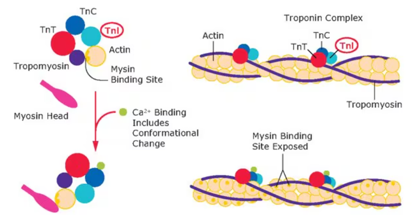
In the resting skeletal muscle myocyte, tropomyosin blocks the myosin binding sites on actin. in muscle contraction the sarcomere shortening by recurrent myosin/actin interactions. During each interaction, the myosin heads work to bring adjacent actin free ends closer to the center of the sarcomere. The complex process leading to muscle contraction, called excitation-contraction coupling
- When a significant motor end plate depolarizing potential overcomes the threshold of the skeletal myocyte, the cell fires an action potential.
- This depolarizing potential is propagated to T tubules, causing a conformational change in the dihydropyridine receptors.
- This conformational change opens ryanodine receptors on the terminal cisternae of the sarcoplasmic reticulum to release Ca from its storage site in the sarcoplasmic reticulum into the intracellular fluid
- Ca2+ binds to troponin C, causing a conformational change in troponin that moves tropomyosin from the myosin binding site on actin. Note: in the presence of Ca2+, troponin C remains in this configuration, leaving the myosin binding site on actin available for myosin interactions.
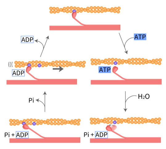
At first, no adenosine triphosphate (ATP) is bound to myosin causing myosin to bind to actin (permanent lack of ATP causes permanent myosin and actin interaction and is the mechanism behind rigor mortis
- ATP then binds myosin causes the release of the actin-myosin cross-link (=myosin disassociated with actin
- Hydrolysis of this bound ATP to ADP plus inorganic phosphate induces a change in myosin to the “cocked position.” Note: only ADP remains attached to myosin.
- The myosin head then interacts with actin at another binding and moving the myosin head toward the positive end of actin (closer to the ends of the sarcomere).
- ADP is released from myosin causing another change that results in the “power stroke.” At this time, myosin is bound to actin and will remain bound without more ATP.
Another ATP molecule binds myosin. The cycle continues, shortening the sarcomere as myosin slides along the actin.
Relaxation occurs when there is a decrease in excitatory motor end plate potentials, a decrease in action potentials and repolarization of the myocyte. Ca2+ gets sequestered back into the SR by Ca2+ -ATPase pumps, decreasing intracellular [Ca2+]. These myocytes also contain Na+/Ca2+ exchangers on the cell surface that use the Na+ electrochemical gradient to exchange Na+ into the cell in exchange for Ca2+ out of the cell. Ca2+ disassociates from troponin C, and tropomyosin blocks the myosin binding sites on actin once again.
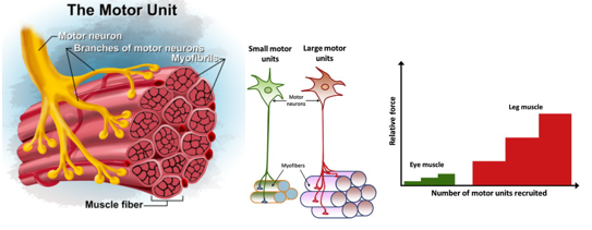
The force of contraction is a summation of the number of motor units recruited and the frequency of action potentials that reach those motor units. The motor unit is defined as a single motor neuron and all the muscle fibers it innervates, with multiple motor units stimulating a single muscle. The number of muscle fibers comprising a motor unit depends on the function of the muscle. Muscles that require fine motor control will involve fewer fibers, whereas larger muscle groups will involve significantly more muscle fibers. Muscles that function over a prolonged period, such as lower back muscles, will asynchronously recruit fibers so that fatigue of individual fibers is spread out over time and space
8) Types of Striated Muscle Contraction
Isometric muscle contraction: A contraction where the muscle keeps the same length despite increasing tension.
Isotonic muscle contraction: A contraction where the tension in the muscle remains unchanged despite a change in muscle length. Isotonic contraction is subdivided into concentric and eccentric muscle contraction.
• Concentric muscle contraction happens when the muscle shorten as it contracts.
• Eccentric muscle contraction happens when the muscle lengthen as it contracts.
9) Muscle Metabolism During Contraction
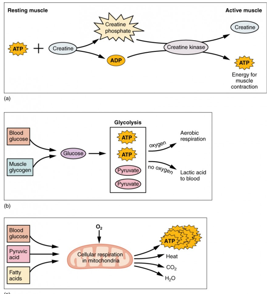
Muscle metabolism is driven by the need to produce ATP for contraction. Muscles first use their small store of ATP, then rely on creatine phosphate for about 15 seconds of extra energy. After this, they obtain ATP from breaking down glycogen and lipids. Without oxygen, glucose undergoes anaerobic glycolysis, producing only 2 ATP and lactic acid, which supports activity for about a minute. With oxygen, aerobic respiration in mitochondria produces up to 36 ATP per glucose and can sustain activity much longer, relying first on muscle glycogen and then on nutrients delivered by the blood. During intense exercise, the heart and lungs must boost oxygen supply. Anaerobic metabolism is fast but inefficient, while aerobic metabolism is slow to start but produces much more ATP.