
skeletal muscle physiology
1) Introduction
Muscle is an organic tissue composed of contractile fibers that produce movement. There are three major muscle types found in the human body: skeletal, cardiac, and smooth muscle. Each muscle type has unique cellular components, physiology and specific functions
2) muscle types
2.1 Skeletal muscle
Skeletal muscle is found throughout the body and functions to contract in response to a stimulus. Skeletal muscle serves many purposes, including producing movement, sustaining body posture and position, maintaining body temperature, storing nutrients, and stabilizing joints. In contrast to smooth and cardiac muscle contraction, most skeletal muscle contraction is under voluntary control, receiving neural inputs allowing conscious control of muscles. Skeletal striated muscle is the most abundant type (over 400 distinct muscles), represents approximately 40% of the human body weight and contains 50 to 75% of all body proteins
2.2 Cardiac Muscle
Cardiac muscle also called the myocardium, like skeletal muscle, is made up of sarcomeres that allow for contractility. However, unlike skeletal muscle, cardiac muscle is under involuntary control. The cardiac muscle is responsible for the contractility of the heart and, therefore, the pumping action. The cardiac muscle must contract with enough force and enough blood to supply the metabolic demands of the entire body.
Cardiac muscle cells (cardiomyocytes) are striated, branched, contain many mitochondria, and are under involuntary control. Each myocyte contains a single, centrally located nucleus surrounded by a cell membrane known as the sarcolemma. The sarcolemma of cardiac muscle cells contains voltage-gated calcium channels, specialized ion channels that skeletal muscle does not possess. Gap junctions between adjacent cardiomyocytes allow for the propagation of coordinated action potentials from one cell to the next in a phenomenon known as electrical coupling
2.3 smooth muscle
Smooth muscle tissue is an involuntary, non-striated muscle found in the walls of hollow organs throughout the body. Smooth muscle is present in all the organ systems below:
Gastrointestinal tract : propulsion of the food
Cardiovascular blood vessel and lymphatic vessels : regulation of blood flow and pressure via vascular resistance
Renal: urinary bladder : regulation of urine flow
Genital : uterus, both male and female reproductive tracts: contractions during pregnancy, propulsion of sperm
Respiratory tract : regulation of bronchiole diameter
Integument : erector pili of the skin
Sensory: the ciliary muscle and iris of the eye :dilation and constriction of the pupil as well as changing lens shape
3) Muscle characteristics
· Excitability is the ability to respond to a stimulus
· contractility is the ability to forcefully shorten
· Extensibility allows muscle tissue to be stretched
· Elasticity is its ability to return to its original length after stretching
4) Somatic motor
Somatic motor are voluntary actions controlled by the somatic nervous system, which governs movement in skeletal muscles. which involve the brain sending signals down through motor neurons in the spinal cord to initiate movements like walking, talking, or grasping an object
5) Macroscopic anatomy of a skeletal muscle
skeletal muscle fibers are made up of endomysium, perimysium, and epimysium, covering the sarcolemma; each muscle fiber is a layer of connective tissue called the endomysium. Capillaries and nerve tissue are present within the endomysium to supply the individual muscle fibers. Multiple muscle fibers join to form fascicles encased by another connective tissue covering known as the perimysium. The perimysium may surround anywhere from 10 to 100 fascicles. Muscle fascicles are further grouped to form a muscle encased by a fibrous tissue envelope called the epimysium.
6) Microscopic anatomy of a skeletal muscle
muscle fibers
Skeletal muscle is a highly organized tissue composed of bundles of muscle fibers called myofibers which are striated, multinucleated cells ranging from 10 to 100 micrometers in diameter and many centimeters long. The oval nuclei are located in the cell's periphery, adjacent to the sarcolemma
Sarcolemma
The sarcolemma is a tubular sheath that encases and defines each muscle fiber, forming a barrier between extracellular and intracellular compartments. The sarcolemma is comprised of a plasma membrane and a polysaccharide coating . Invaginations within the sarcolemma are termed transverse tubules (T tubules), which function as a major location for ion exchange.
sarcoplasm
The sarcoplasm of a muscle fiber is comparable to the cytoplasm of other cells, but it contains significant reserves of glycogen as well as myoglobin, a protein (red pigment) that constitutes an oxygen reservoir inside the muscle cell and does not exist in any other type of cell. Muscle cells contain the usual organelles as well as highly modified organelles, namely the myofibrils and the sarcoplasmic reticulum.
Myofibrils
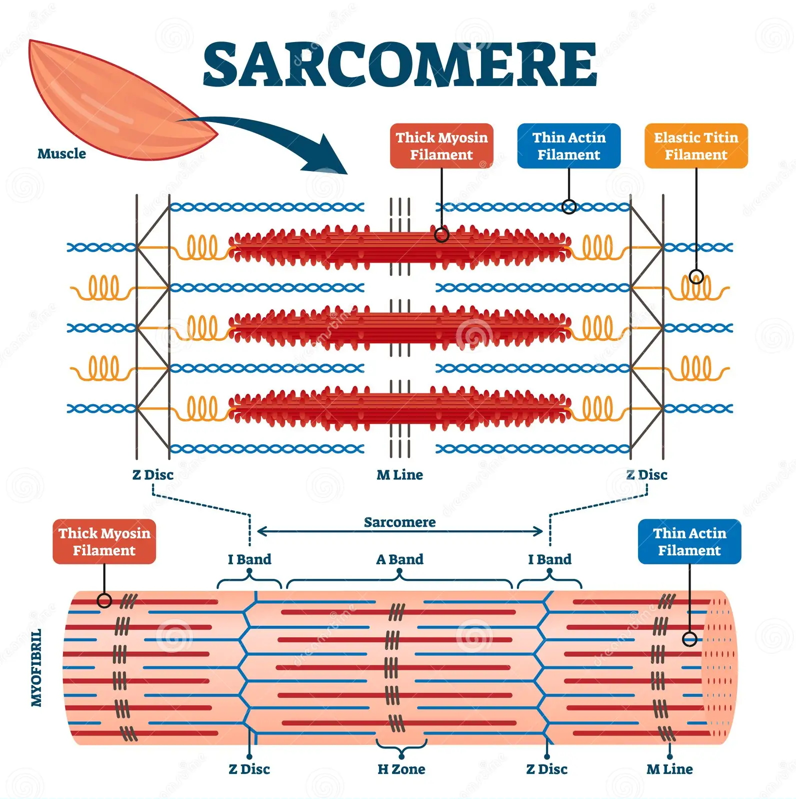
Each muscle fiber is composed of several hundred to several thousand myofibrils. Myofibrils are composed of actin (thin filaments), myosin (thick filaments), and support proteins. The arrangement of actin and myosin gives skeletal muscle its microscopic striated appearance and creates functional units called sarcomeres. When viewed under electron microscopy, sarcomeres are arranged longitudinally and include the M line, Z disk, H band, A band, and I band.
Sarcomeres
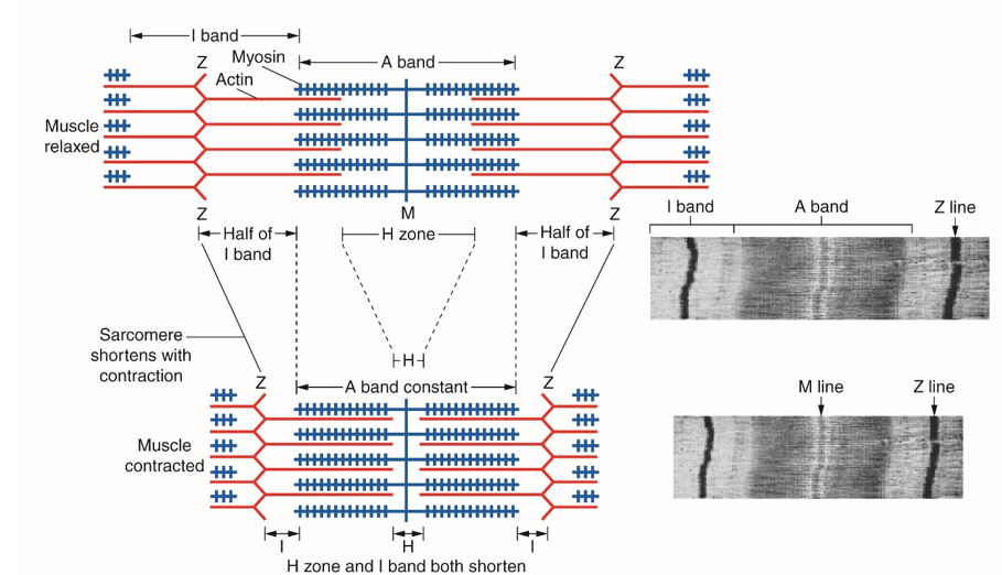
The Z line, or Z disk, is the terminal boundary of the sarcomere, where alpha-actinin acts as an anchor for the actin filaments. The M line is the central-most line of the sarcomere, where myosin filaments are anchored together through binding sites within the myosin filament. The H band contains the M line and is the central region of the sarcomere that contains only myosin filaments. The A band is a larger portion of the sarcomere that contains the entirety of the myosin fibers and includes regions of actin and myosin overlap. The I band covers the terminal regions of two adjacent sarcomeres and contains only actin filaments. The H and I bands shorten with muscle contraction, while the A band remains a constant length.
Actin
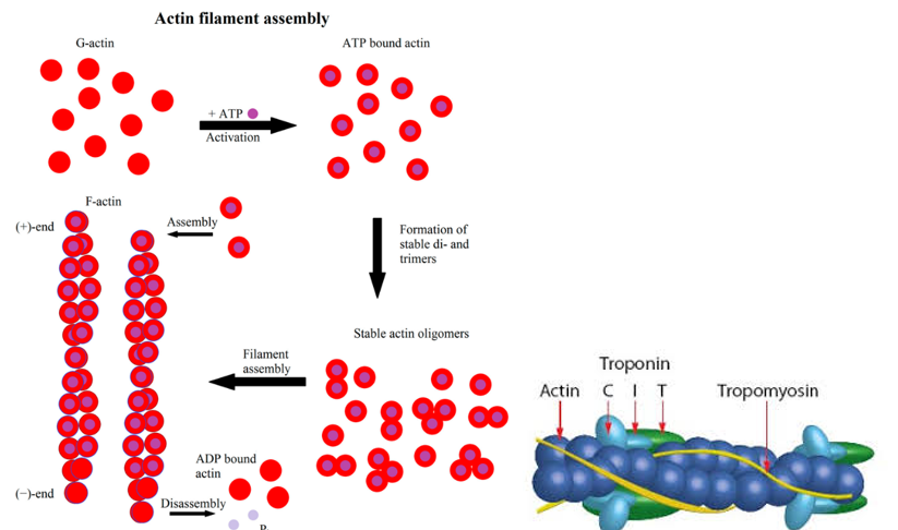
Actin filaments are double-helical structures, known as filamentous-actin (F-actin), composed of monomeric G-actin units. G-actin exhibits polarity and creates a positive and negative end within the sarcomere, with the positive end situated toward the terminal end of the sarcomere. Tropomyosin is a helical protein that runs along the actin double helix within its groove. Troponin binds tropomyosin by a troponin complex at every seven-actin monomer and is composed of troponin-C (Tn-C), troponin-I (Tn-I), and troponin-T (Tn-T). Tn-T binds tropomyosin, Tn-I inhibits actin and myosin binding, and Tn-C binds calcium.
Myosin
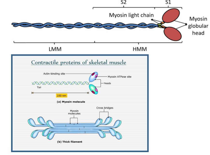
Myosin proteins are composed of two regions: light meromyosin and heavy meromyosin. Light meromyosin (LMM) binds other light meromyosin regions to anchor myosin at the M line. Heavy meromyosin (HMM) is further subdivided into two regions; the S-1 portion, or the myosin head, binds actin and contains an ATPase portion, while the S-2 portion is the location of the power stroke.
Support proteins
Support proteins within the sarcomere include titin, desmin, myomesin, C protein, nebulin, and plectin. Plectin tethers the Z disks of adjacent myofibrils to each other. Desmin helps maintain myofibril alignment, connects to the cytoskeleton and other structural elements within the cell, and distributes contractile force. Myomesin and C protein are myosin-binding proteins that function to tether and stabilize myosin at the M line. Titin is found at the Z disk and anchors myosin longitudinally within the sarcomere. nebulin ensuring thin filaments are the correct length