Introduction
The nervous system is divided into the central nervous system (CNS) and the peripheral nervous system (PNS). The CNS includes the brain and spinal cord, while the peripheral nervous system consists of all the neural tissue outside of the brain and spinal cord. The PNS includes the cranial nerves and spinal nerves, sensory receptors and ganglia (sensory nerve pathways and voluntary and involuntary motor pathways) . The CNS's responsibilities include receiving, processing, and responding to sensory information.
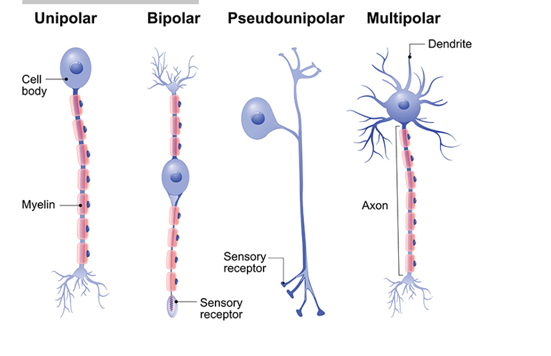
Neurons: There are many billions of nerve cells, also called neurons, in the nervous system. The brain alone has about 100 billion neurons in it. Each neuron has a cell body and various extensions. The shorter extensions called dendrites receive signals from, other neurons and pass them on to the cell body. The signals are then passed on via a long extension (the axon), which can be up to a meter long
Electrophysiology of cell membrane
All cells have an intracellular chemical composition that differs from the extracellular environment, creating a potential difference across the membrane. In most cells (non-excitable cells), this transmembrane potential difference remains relatively stable. However, in excitable cells (such as nerve and muscle cells), this potential can change rapidly, shifting from a resting state (resting potential) to an active state characterized by an action potential (AP), before quickly returning to its equilibrium state..
Resting potential
The resting potential (RP) corresponds to a potential difference (PD) or voltage difference, expressed in millivolts (mV), across the cell membrane. The RP establishes an electrical polarity in which the extracellular environment is positive (+) and the intracellular environment is negative (−). This electrical polarity creates an electrical force gradient, known as the electromotive force (EMF), which influences the movement of ions across the cell membrane. The RP results from the unequal distribution of ions on either side of the cell membrane (CM), particularly Na⁺ and K⁺. This distribution is maintained by the semi-permeable nature of the membrane, which allows limited diffusion of K⁺ and even less diffusion of Na⁺ through leak channels. The activity of the Na⁺/K⁺ ATPase pump also helps preserve this imbalance by counteracting the passive diffusion of Na⁺ and K⁺.
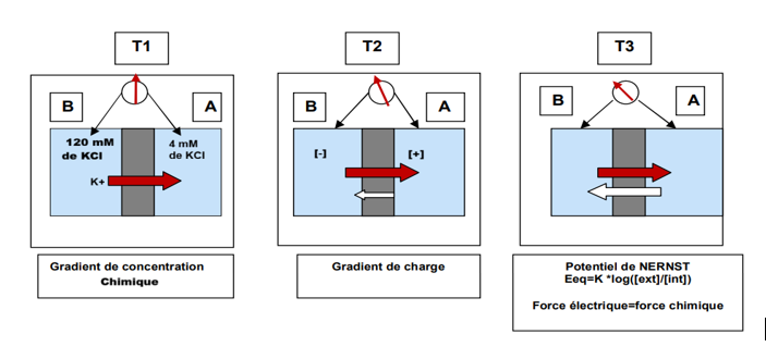
The potential difference measured at equilibrium for a given ion is called the equilibrium potential of that ion. The equilibrium potential therefore corresponds to the electrical gradient which, opposing the chemical gradient, maintains the ion at equilibrium. It is calculated using the Nernst equation
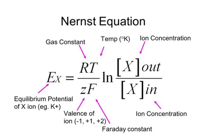
R = 8,28 J/mol/°K
T = 273 + temperature in °C
F = 96000 Coulombs
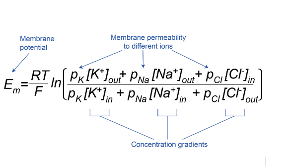
However, the axonal membrane is permeable to several ions (Na⁺, K⁺, and Cl⁻). Therefore, its resting potential depends on the equilibrium potentials of each ion as well as the relative permeability of the membrane to these ions. The Nernst equation, which applies to a single ion, cannot be used to calculate the resting membrane potential. For this reason, the Goldman equation is used, as it considers the relative permeabilities of the membrane to the different ions.
the measurement of the resting potential
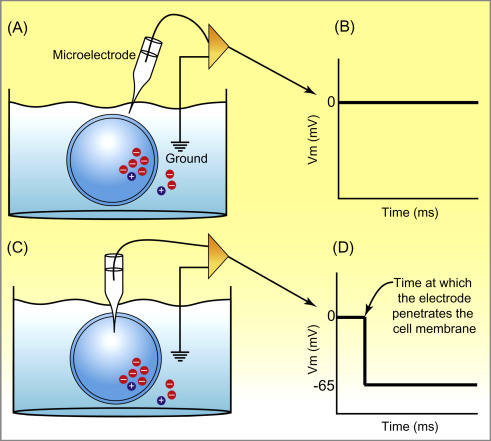
The measurement of the resting potential is performed using the intracellular microelectrode technique. The objective is to record the potential difference that exists across the plasma membrane ; therefore, it is necessary to measure and compare the intracellular potential with the extracellular potential.
To measure the resting membrane potential of a cell, the two terminals of a voltmeter are connected: one to a reference electrode and the other to a micropipette. The tip of the micropipette is brought close to the cell surface, and the reference electrode is placed in the extracellular solution. The signal is amplified and transmitted to an oscilloscope. At this stage, the voltmeter shows no potential difference (0 mV), then the tip of the micropipette penetrates the cell, the voltmeter displays the potential difference between the intracellular and extracellular environments. The measured value corresponds to the resting potential (e.g., −75 mV), meaning that the intracellular potential is 75 mV lower than the extracellular potential. The resting potential varies depending on the cell type:
– Smooth muscle cells: −60 to −40 mV
– Skeletal muscle cells: −90 mV
– Cardiac cells: −80 mV
Action potential
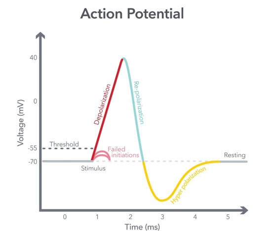
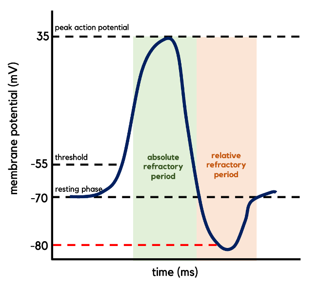
The basic unit of information transmission at the neuronal level is a brief electrical impulse that travels along the axon, called an action potential. This is a temporary reversal of the membrane potential, generated by an extracellular stimulus of sufficient intensity (threshold value), and results from the opening and closing of various voltage-gated ion channels.
This potential, which corresponds to a temporary change in membrane polarity, consists of three phases:
Depolarization: Extremely brief, lasting only a fraction of a millisecond, it results from an increase in sodium (Na⁺) permeability, allowing an influx of positive charges. Consequently, the membrane potential becomes less negative, typically changing from about −60 mV to +40 mV.Repolarization: Slightly slower, this phase allows the membrane potential to return toward its resting level. It corresponds to the closure of sodium channels and the opening of potassium channels.
Hyperpolarization: Even slower (lasting several milliseconds) and of low amplitude, this phase corresponds to an excessive efflux of potassium during repolarization, making the membrane potential more negative than the resting potential.
These phenomena are mediated by the function of voltage-gated ion channels.
The time between hyperpolarization and the resting state:
potassium channels are not immediately inactivated when the fiber returns to its resting potential, potassium ions continue to leave the cell, causing a slight hyperpolarization until potassium permeability returns to its resting value K+ (channels will close progressively). At the same time, the Na⁺/K⁺ pump becomes active and expels the sodium ions that entered during the depolarization phase
Action potential stimulation and measurement
Imposed potential:
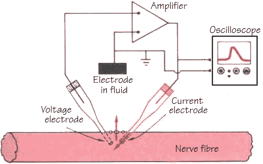
Two microelectrodes are implanted in a cell. One is used to measure the
membrane potential relative to a third, indifferent electrode placed in the
extracellular environment. The other intracellular electrode is used to inject
an electric current.
If an electric current is passed through the cell, adding positive charges, the membrane potential becomes less negative (depolarization). Conversely, if the electric current flows in the opposite direction, the membrane potential becomes more negative (hyperpolarization)
Patch Clamp:
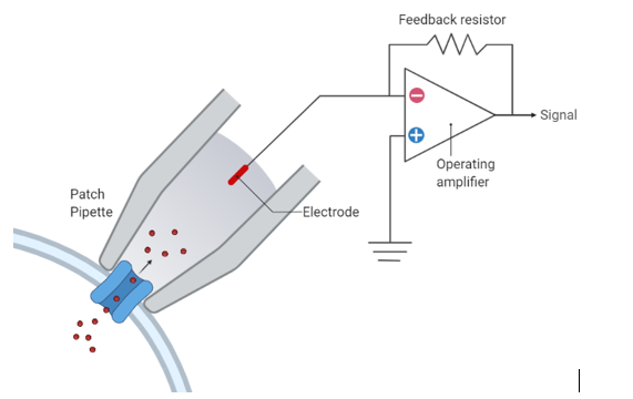
This technique allows the study of ion channel function at the single-channel level. A patch of membrane is isolated inside a glass micropipette with a tip diameter of approximately 1 µm. Using appropriate electronic equipment, a controlled potential is applied to the membrane patch, and the resulting ionic currents flowing through the isolated channel(s) are recorded
Ion channel inhibitor method:
Adding tetrodotoxin (TTX) to the preparation prevents an action potential from occurring. TTX has the unique property of blocking sodium channels, thereby preventing depolarization.
Conversely, adding tetraethylammonium (TEA) to the preparation causes repolarization to occur much later once the fiber has depolarized. This is because TEA blocks potassium channels, so the fiber must wait until the sodium channels are completely inactivated and the Na⁺/K⁺ pump restores the initial ionic concentrations before repolarization can take place.
Excitability and conductibility
Excitability and conductivity are two inseparable properties of the neuron. The former allows it to generate an action potential following a suprathreshold stimulation, and the latter allows it to propagate. However, at the moment the action potential is generated, the fiber is depolarized, making it impossible to depolarize it again. It is therefore necessary to wait until the membrane potential returns to a value below the critical threshold before the neuron can be excited again. This leads us to distinguish two periods that characterize its excitability:
An absolute refractory period (ARP) during which any stimulation, even suprathreshold, is ineffective because the fiber is already depolarized.
• A relative refractory period (RRP) during which a second action potential can be generated, provided that the depolarization produced by the stimulus reaches the critical threshold. This requires a stronger stimulus, as the resting potential has not yet been fully restored.
In the body, an action potential can arise in two ways:
• First case: If a stimulus reaches a sufficient intensity, it produces a local change in potential, called a receptor potential and/or generator potential. When this potential exceeds a critical threshold, it triggers an action potential in the nerve fiber originating from the receptor. This action potential is then transmitted to the brain.
Second case: Synaptic contacts allow a neuron to receive multiple signals from other neurons, resulting in either depolarizations (excitatory synapses) or hyperpolarizations (inhibitory synapses). These postsynaptic potentials (EPSPs and IPSPs) propagate to the cell body, where an algebraic summation of depolarizations and hyperpolarizations occurs. If depolarization predominates and reaches the threshold, an action potential is generated at the axon hillock (trigger zone)
The spike (action potential) follows the all-or-none law, meaning it occurs only when the stimulus reaches a threshold: a spike is generated if the stimulus intensity is at or above the threshold, and no spike occurs if the stimulus is below the threshold. Once it occurs, the action potential immediately reaches its maximum amplitude, and this amplitude does not increase further, even with suprathreshold stimulus intensities.
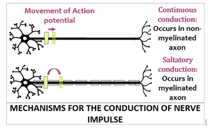
Continuous Conduction: It refers to the non-myelinated conduction, where the flow of nerve-impulse is slower (0.1 m/s). It occurs in unmyelinated axons, where the ions flow throughout the axon segment via voltage-gated channels.
Saltatory Conduction: It refers to the myelinated conduction, where the action potential plunges much faster (100 m/s) from one node to another. It occurs in myelinated axons, where the flow of ions is discontinuous because of the uneven distribution of voltage-gated channels