
Ligand-receptor Interaction
1. Introduction
Intercellular communication is one of the characteristics of multicellular organisms, is done by the secretion of chemical signals or ligands released by secretory cell to target cell has ligand-specific receptors. Allows cell survival, proliferation, and/or differentiation. The absence of signals leads to cell death.
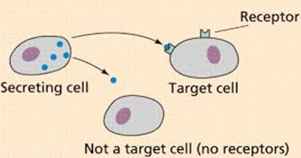
2. type of communication
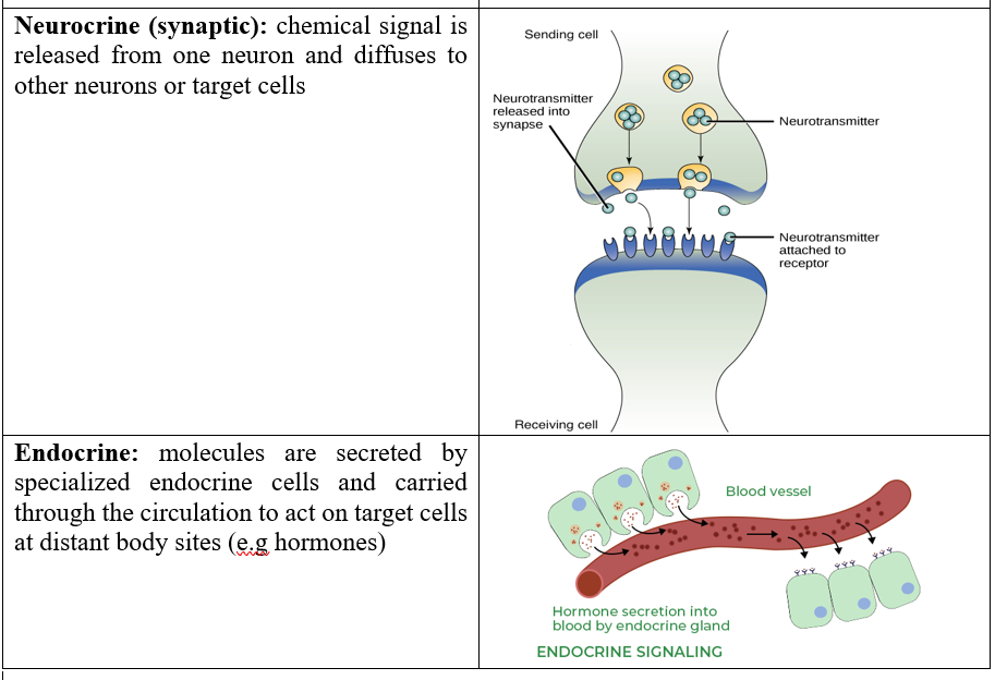
Direct :
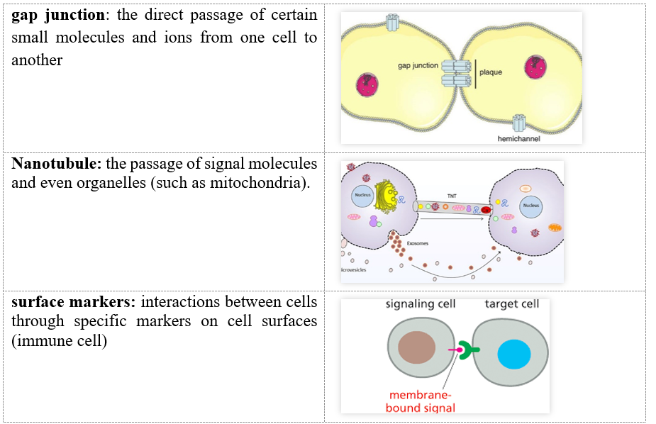
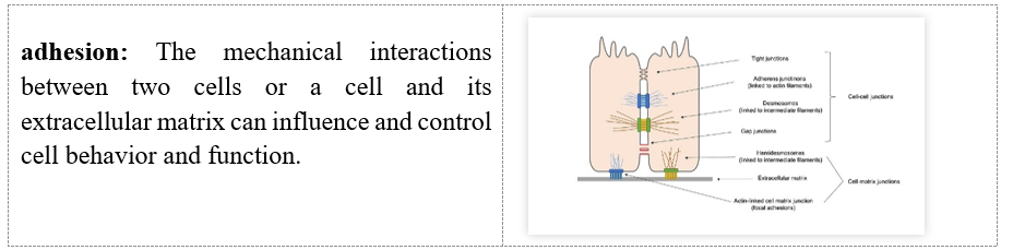
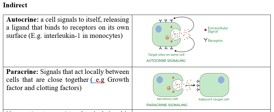
3. ligand:
water-soluble: eg Neurotransmitters, Peptide hormones, Growth factors, prostaglandins bind to cell surface receptors
Fat-soluble: eg steroid hormone, thyroxine, retinoic acid, bind to intracellular receptors
4. Intercellular communication :
Reception Phase: Consists of the detection and recognition of the ligand and its binding to the specific receptor.
Transduction Phase: a cascade of interactions between several intermediary molecules (transduction pathway).
Cellular Response Phase: generation a cellular response, i.e., changes in cellular behavior
5. Receptors:
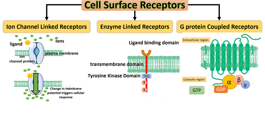
Surface receptors
Intracellular receptors
5.1 Surface receptors:
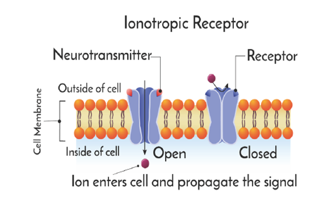
a. Ionotropic receptor
These are polymeric receptors, generally pentameric, formed of 5 distinct protein subunits (α, β, γ, δ) which surround an ion channel, one or more of which have sites capable of binding the ligand, generally the α subunit.
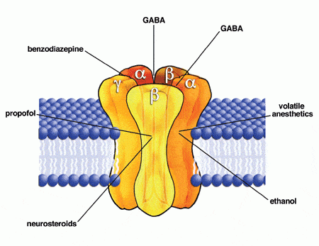
The binding of a mediator molecule to its site causes a change in the conformation of the channel protein, and the opening of the ion channel and diffusion of ions across the membrane. Ionotropic receptors are involved in both excitatory and inhibitory synapses e.g nicotinic cholinergic receptor (acetylcholine receptor channel).
b. G protein-coupled receptors (Metabotropic receptor)
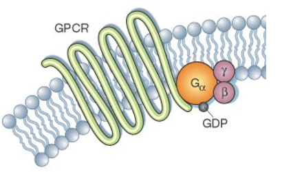
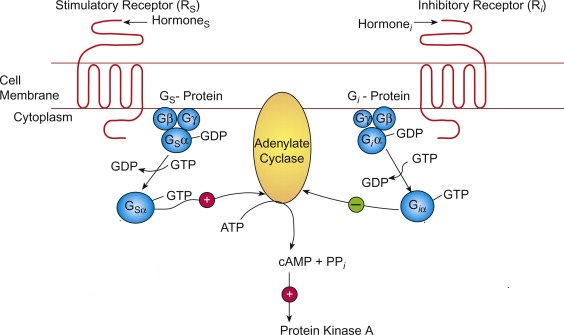
G protein-coupled receptors (GPCRs) are integral membrane proteins containing an extracellular amino terminus, seven transmembrane α-helical domains, and an intracellular carboxy terminus; GPCRs were named for their common ability to associate with heterotrimeric G proteins (Gαβγ). The α subunit has a GDP/GTP binding site.
The binding of the ligand (first messenger) to its specific GPCR triggers a series of reactions:
- The receptor changes its three-dimensional conformation and activates the G protein;
-when a GPCR is activated, it induces the α subunit to release GDP and instead binds to guanosine triphosphate (GTP).
-The α subunit is activated by binding to GTP and detaches from the β,γ dimer of the G protein;
-The α subunit GTP activates an effector (an ion channel or an enzyme)
-The α subunit of G protein has a GTPase activity, and once it hydrolyzes GTP to GDP, the α GDP molecule reassociates with the β-γ dimer and becomes inactive.
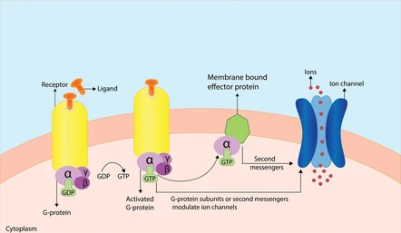
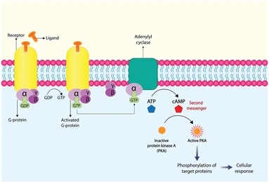
In some cells, the G protein directly activates an ion channel, causing the passage of ions. In other cells, the activated G protein activates a membrane enzyme, such as adenylate cyclase (effector), which forms small molecules called cAMP (cyclic adenosine monophosphate), which act as a second messenger. The cAMP activates a second intracellular enzyme called protein kinase A (PKA), which amplified phosphorylation cascade leads to the regulation of gene expression or opening of the ion channel
in the case of the αi-GTP complex the result will be the inhibition of the formation of the second messenger
Guanylate cyclase is an enzyme that catalyzes the conversion of GTP to cyclic GMP (cGMP), an intracellular second messenger
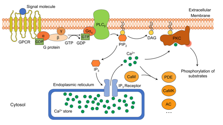
Many GPCRs elicit their physiological function by activating an inositol phospholipid signaling pathway via phospholipase C-β (effector) (PLCβ). This particular pathway is mediated by a sub-class of GPCRs often coupled to a G proteinα q (Gαq) that leads to the activation of PLCβ. Activated PLCβ can hydrolyze phosphoinositol bisphosphate (PIP2) to form-diacylglycerol (DAG) and inositol trisphosphate (IP3) (second messenger) While IP3 is known to interact with the IP3 receptors at the endoplasmic reticulum (ER) and results in intracellular calcium release, DAG is best known for its role in the activation of protein kinase C (PKC).
c. Enzyme receptors (tyrosine kinase receptors)
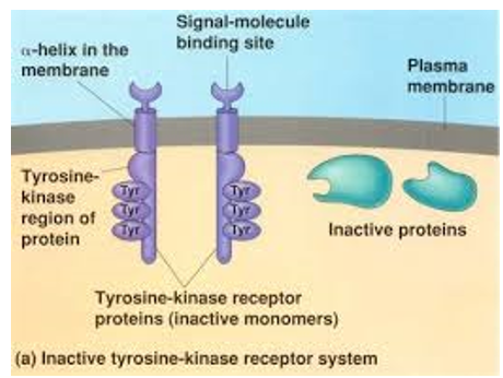
The receptor itself has an enzymatic activity. Its activation by the primary messenger, this activity which is of the kinase or phosphatase type. Most of them are receptor tyrosine kinases (RTK), one of the best known being the insulin receptor
The insulin receptor is composed of two α subunits and two β subunits.2 extracellular α subunits: These are involved in the binding and recognition of insulin. 2 transmembrane β subunits: These carry the catalytic activity generated by the hormone/receptor binding. A disulfide bridge links each α unit to a β
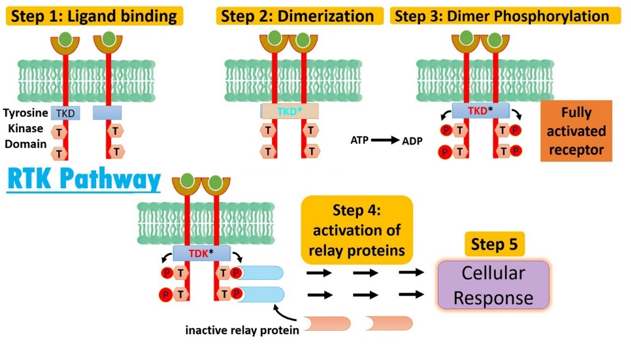
This process begins when insulin binds to the receptor alpha subunit in the extracellular domain. The conformational change of the receptor, the monomeric units of receptors come together and dimerises. Dimerization activates tyrosine kinase in the intracellular domain of the receptor undergoes autophosphorylation (an -OH group carried by a tyrosyl residue becomes -O-P) and phosphorylation of the IRS (insulin receptor substrate) adjacent to the receptor (R-OH R-O-P)which transmit the hormonal message through a cascade of phosphorylation/dephosphorylation reactions that will cause the observed biological effects (formation of glucose transporters)
5.2 Intracellular receptors
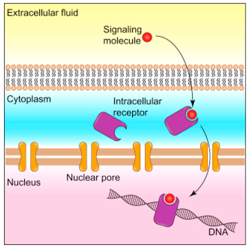
Intracellular receptors, also known as nuclear receptors, regulate cellular functions by altering gene expression. These receptors are classified into 2 types—type I and type II nuclear receptors (intranuclear and extranuclear) activated by lipophilic hormones such as: thyroid (thyroxine T4, triiodothyronine T3) and steroid (estrogen, progestin).
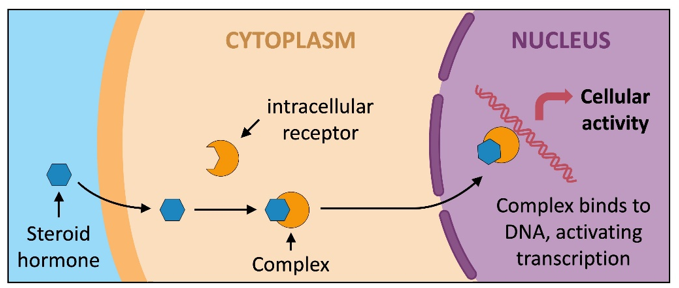
a. Type I intracellular receptors: In the inactive state, type I nuclear receptors are found in the cytoplasm and are usually bound to a chaperone protein. Following the binding of a ligand, the receptor undergoes a conformational change that releases the chaperone protein and allows the receptor to translocate to the nucleus. Once in the nucleus, the ligand-receptor complex binds to a specific DNA sequence and acts as a transcription factor to modulate the transcription of one or more genes. Type I intracellular receptors include the glucocorticoid, androgen, and progesterone receptors.
b. Type II intracellular receptors: Type II intracellular receptors are located in the nucleus regardless of ligand-binding status. In the inactive state, these receptors are bound to co-repressor proteins. The co-repressor proteins are released upon ligand binding, allowing the receptor to bind to DNA and modulate gene expression. Type II intracellular receptors include retinoic acid and thyroid hormone receptors
6. Signal amplification
The second messenger activation steps play an important role in amplifying the initial signal. Binding a single ligand to a receptor activates several adenyl cyclase (AC) molecules (e.g., 10), each of which activates several (e.g 100) cAMP molecules. Each cAMP molecule then acts on a single protein kinase A, which phosphorylates and thus influences many (e.g 100) specific proteins, such as enzymes. Each enzyme, in turn, is responsible for producing many molecules of a particular product; the result of this cascade is a huge amplification of the initial signal. In this way, very low concentrations of ligands can trigger a very significant response in the cell.
7.Message Interruption
Catabolism of Second Messengers
- Ca++: Four Mechanisms
* Expulsion of Ca++ to the outside by a Ca++-dependent ATPase pump;
* Reuptake of Ca++ by the sarcoplasmic reticulum using an ATPase pump;
* Reuptake by the mitochondria;
* Calcium-sodium system that releases Ca++ to the extracellular environment.
- cAMP: Degraded by phosphodiesterase,