
Structure of the cell membrane
Structure and composition of the
plasma membrane
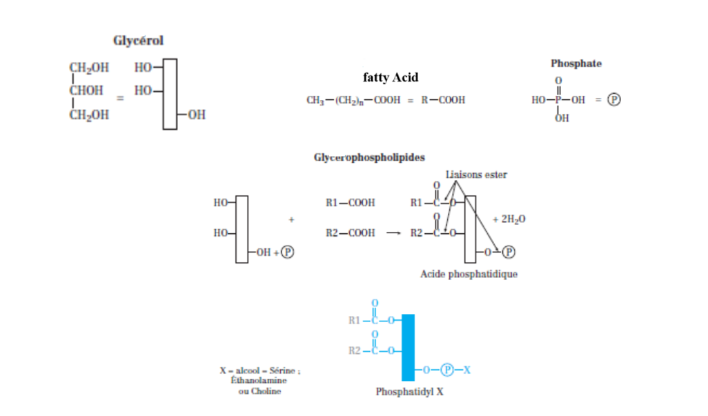
Three types are distinguished:
1) Glycerophospholipids (the most abundant).
They are classified as follows:
• Phosphatidylserine (PS)
• Phosphatidylethanolamine (PE)
• Phosphatidylcholine (PC) = lecithin
Glycerophospholipids provide the membrane fluidity necessary for many cellular functions associated with the plasma membrane, such as communication, transport, movements, and adhesion
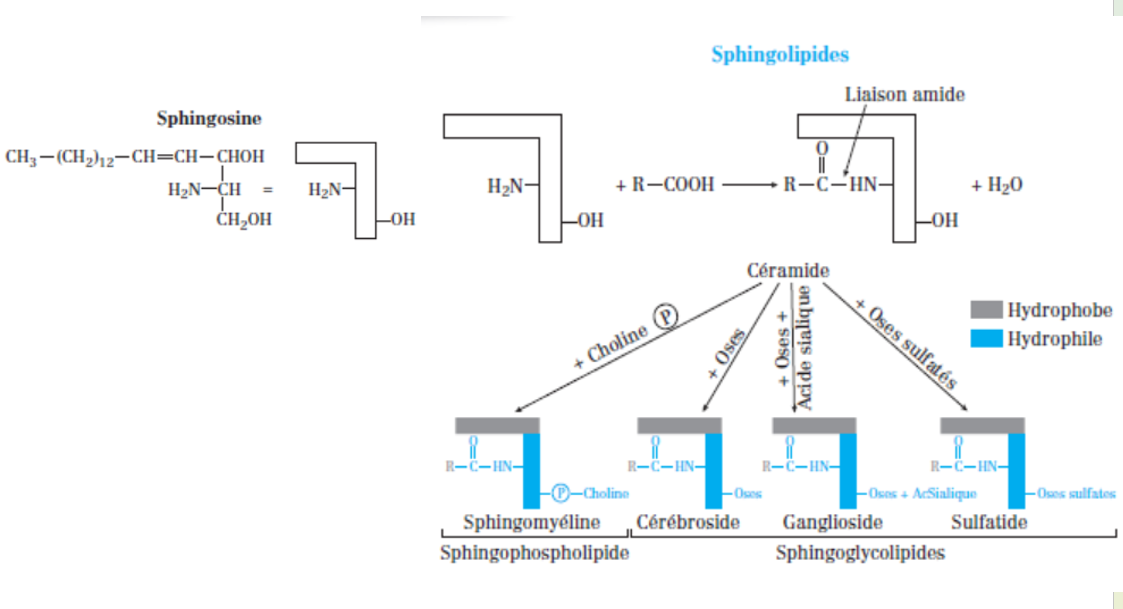
Sphingolipids
Difference from glycerophospholipids: glycerol is replaced by sphingosine. They
are classified as follows:
• Ceramide =
sphingosine + fatty acid
• Sphingophospholipids
=
sphingosine + fatty acid + phosphate + alcohol. The most well-known is sphingomyelin
(where the alcohol is choline).
• Sphingoglycolipids,
including:
– Ganglioside
=
ceramide + sugars + sialic acid
– Cerebroside =
ceramide + sugars
– Sulfatide =
ceramide + sulfated sugars
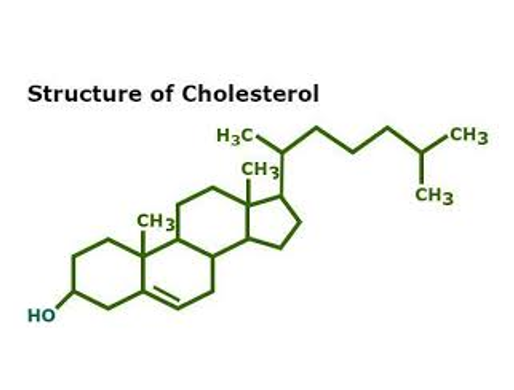
Cholesterol: is amphiphilic: hydrophilic due to its –OH group, and hydrophobic due to its four carbon rings and to its aliphatic side chain, Cholesterol regulates membrane fluidity: it stiffens the membrane at high temperature and increases its fluidity at low temperature.
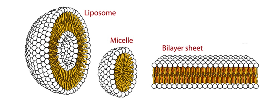
When membrane lipids are in an aqueous phase, they can organize in several different ways:
c) Liposome: These are artificial structures, produced in vitro. Liposomes have the shape of small spherical vesicles delimited by a lipid bilayer and filled with an aqueous medium. They can be used as vectors to deliver drugs to cells, as they could fuse with the plasma membrane to release their contents.
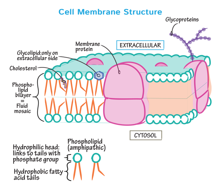
They have a hydrophobic part and a hydrophilic part. This property allows them to organize into a bilayer that embeds intrinsic proteins.
– Outer leaflet: Sphingomyelin, Phosphatidylcholine, Glycolipids
– Inner leaflet: Phosphatidylethanolamine, Phosphatidylserine
C) Fluidity of the plasma membrane
Membrane
fluidity depends on several factors:
• The
nature of the fatty acids that make up the phospholipids:
– Unsaturated fatty acids increase fluidity, while saturated fatty acids make
the plasma membrane more rigid.
– The longer the carbon chain of these fatty acids, the more rigid the membrane
becomes.
• The amount of cholesterol: Membrane fluidity decreases as cholesterol content increases.
• Temperature: Fluidity increases as temperature rises.
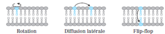
d) Movements of lipids
Three types of movements are possible for lipids:
– Rotation on themselves
– Movement within the same leaflet: lateral diffusion
– Transfer from one leaflet to the other: flip-flop or transverse diffusion.
Lipid flip-flop requires the intervention of enzymes: flippases
(which need energy in the form of ATP)
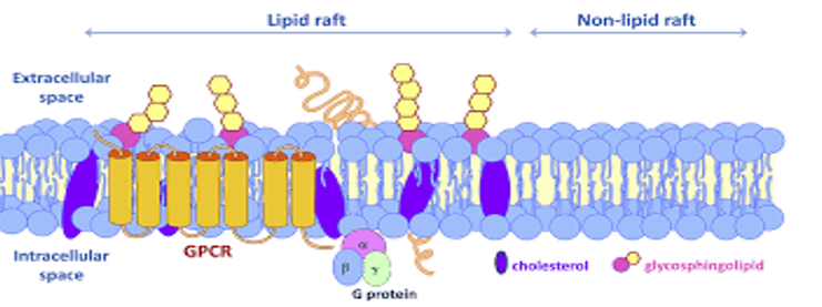
e) Lipid rafts
These are lipid microdomains, also called 'rafts' or DIGs (Detergent-Insoluble Glycolipid-enriched microdomains).specialized domains within cell membranes, enriched in cholesterol and sphingolipids, which serve as platforms for organizing proteins and lipids to regulate various cellular processes
Membrane proteins
– rotation;
– lateral diffusion
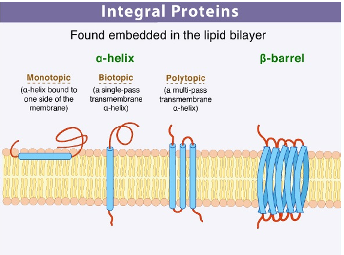
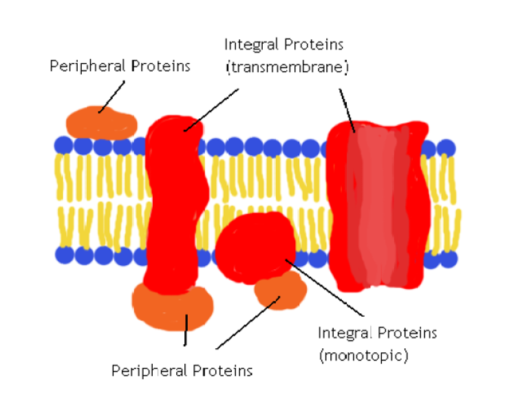
a) Transmembrane proteins: They are very tightly associated
with the membrane and therefore very difficult to extract. For their
purification, detergents or organic solvents are used.
• They span the membrane once or several times. When they span it once, they
are called bitopic;
when they span it multiple times, they are called polytopic.
Interactions between membrane lipids and transmembrane proteins are
non-covalent and related through the hydrophobic amino acids of the protein.
Transmembrane domains generally extend over about twenty amino acids and are
often organized as α-helices. The extracellular and cytosolic domains of these
proteins are usually hydrophilic.The
extracellular parts of the protein may be glycosylated. There are some
transmembrane proteins that do not span the membrane completely. These are
referred to as monotopic proteins.
b) Anchored proteins: These proteins are associated with membrane lipids through covalent bonds. Some
proteins are linked to the inner leaflet of the plasma membrane via:
– a fatty acid: these are acylated proteins;
– a fatty alcohol: these are prenylated proteins.
There are also proteins anchored to the outer leaflet via phosphatidylinositol. These proteins are called glypiated or anchored by GPI (Glycosyl Phosphatidyl Inositol).
2) Extrinsic (or peripheral) proteins
These proteins can be found on the extracellular or cytosolic side.
They are never covalently bound to the lipid bilayer; they form weak interactions (hydrogen or ionic bonds) with:
– the polar head groups of membrane lipids;
– the polar regions of intrinsic proteins.
These proteins are purified by simply changing the pH or ionic strength and can be glycosylated.
Glycocalix or cell coat
– cell recognition and identity;
– their adhesion to their environment.
a) Blood Groups
The specificity of red blood cell membrane antigens depends on the nature of the constituent oligosaccharides. These oligosaccharides are carried by membrane glycolipids.
b) The Major Histocompatibility Complex or MHC
Glycoproteins of nucleated cells. They are:
Adhesion linked to CAMs (“cell adhesion molecules”) = membrane glycoproteins
These carbohydrates play a role in:
• Cell migration.
• Cell/cell adhesion.
• Cell/extracellular matrix adhesion.