
Chapter I : The membrane ultrastructure (part 2)
I.5.1.5.Glycolipids
Glycolipids are of two types: glyceroglycolipids and sphingoglycolipids.
► Glycolipids are always associated with the outer leaflet of the membrane and belong to a structure known as the glycocalyx. The glycocalyx is the carbohydrate-rich peripheral zone of the cell.
► Glycolipids result from the linkage of a single hexose or an oligosaccharide to a hydroxyl group belonging either to glycerol (in glyceroglycolipids) or to sphingosine in a sphingolipid.
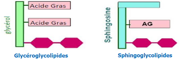
Figure 11 : glyceroglycolipids and sphingoglycolipids.
lipid self-assemble
Phospholipids can self-assemble in an aqueous environment to form liposomes, micelles, or lipid bilayers.
v Micelles: These are typically spherical aggregates where the single-tailed lipid molecules are wedge-shaped (inverted cone). The hydrophobic tails point inward to form a core that excludes water, and the hydrophilic heads form the outer surface in contact with the aqueous solution.
v Lipid Bilayers: These are bimolecular sheets where the phospholipids are cylindrical in shape, meaning the cross-sectional area of the head group is approximately equal to that of the two tails. The tails of two layers point toward each other, forming a hydrophobic interior core sandwiched between two layers of hydrophilic heads facing the water.
v Liposomes: When a lipid bilayer sheet closes on itself to avoid exposed hydrophobic edges, it forms a sealed, spherical vesicle enclosing an internal aqueous compartment. Liposomes can be unilamellar (a single bilayer) or multilamellar (multiple concentric bilayers separated by water layers).
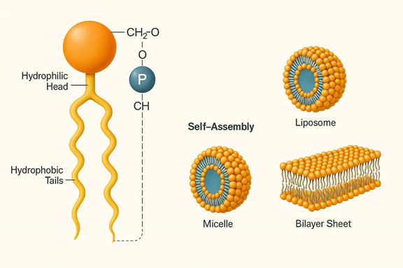
Figure12 : lipid self-assemble
I.5.2. Membrane Proteins
Membrane proteins either integrate into the lipid bilayer or associate with its surface. The cell membrane, or plasma membrane, is a lipid bilayer that encloses the cell, acting as a barrier and offering protection. These proteins exhibit both hydrophilic (water-attracting) and hydrophobic (water-repelling) properties, which enable their stable integration and function within the phospholipid bilayer.
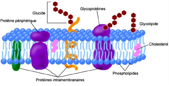
Figure 13 : Proteins are at the origin of membrane dynamics.
Hydrophilic Regions: These polar parts of membrane proteins are usually found on the protein's surface, where they can interact with water molecules or the polar head groups of membrane lipids.
Hydrophobic Regions: Comprising non-polar amino acid residues, these regions often form transmembrane α-helices or β-barrel structures that integrate into the hydrophobic core of the phospholipid bilayer, avoiding interaction with water.
I.5.2.1.Types of Membrane Proteins
Membrane proteins can be broadly categorized into three groups based on their location and interaction with the lipid bilayer:
· Integral Membrane Proteins (Intrinsic Proteins)
· Peripheral Membrane Proteins (Extrinsic Proteins)
· Lipid-Anchored Proteins
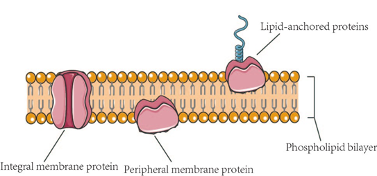
Figure 14 : Membrane Proteins
A-Integral Membrane Proteins
Integral membrane proteins, or intrinsic proteins, embed themselves within the phospholipid bilayer and are stabilized by interactions between their hydrophobic amino acid residues and the hydrophobic phospholipid tails. These proteins are essential for various cellular functions, including transport, signaling, and structural integrity.
A key subset of these proteins is transmembrane proteins, which extend across the lipid bilayer and have sections exposed on both the interior and exterior surfaces of the cell membrane. They typically comprise one or more alpha-helices or beta-barrels, which interact with the membrane's hydrophobic core, while their hydrophilic regions protrude into the aqueous environments inside and outside the cell.
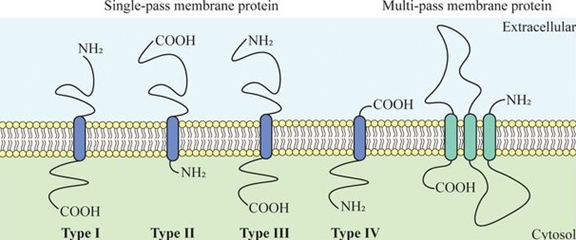
Figure 15: Several types of transmembrane proteins.
B- Transmembrane Proteins
Transmembrane proteins typically have one or more alpha-helices or beta-barrels that span the membrane. Alpha-helical proteins are more common and can function as channels or receptors. The hydrophobic regions of these proteins interact with the membrane's hydrophobic core, while hydrophilic regions protrude into the aqueous environments inside and outside the cell.
These proteins span the phospholipid bilayer with one (single-pass) or several (multipass) segments. Single-pass membrane proteins can be further categorized into various types, as classified in UniProt.
Type I: N-terminus on the extracellular side with the signal peptide removed.
Type II: N-terminus on the cytosolic side, with the transmembrane (TM) domain near this terminus acting as an anchor.
Type III: N-terminus on the extracellular side but lacking a signal sequence.
Type IV: N-terminus on the cytosolic side, with the TM domain near the C-terminus serving as an anchor.
C-Peripheral Membrane Proteins
Peripheral membrane proteins, also known as extrinsic membrane proteins, are proteins attached to the inner and outer surfaces of the cell membrane, making up 20-30% of all membrane proteins. Unlike integral membrane proteins, which are amphipathic and embedded within the lipid bilayer (with some spanning the entire bilayer and exposed on both sides, known as transmembrane proteins), peripheral membrane proteins do not penetrate the lipid bilayer. Instead, they bind to the polar head groups of lipid molecules or to the hydrophilic regions of transmembrane proteins through ionic bonds. These proteins are entirely exposed on either side of the lipid bilayer and are only indirectly associated with the membrane.
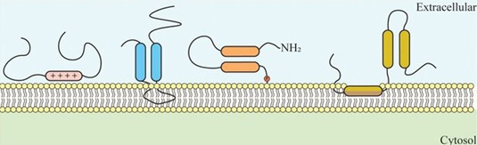
Figure 16 : Schematic representation of peripheral proteins.
I.5.2.2.Functions of Membrane Proteins
Although their phospholipid bilayer determines the structure of any biological membrane, membrane proteins perform most of their functions. Some of the most critical functions performed by membrane proteins are:
1. Enzymatic Functions: Some membrane proteins act as enzymes with their active sites exposed to their substrate molecules. Sometimes, several enzymes in a membrane organize as a team, carrying out sequential steps of a metabolic pathway.
2. Transportation: Some proteins are responsible for facilitated diffusion and active transport. During facilitated diffusion, the membrane-spanning proteins provide a hydrophilic channel that allows the passage of particular solutes. In contrast, others work by a conformational change. Some of these proteins use ATP to actively pump substances across the membrane, thus helping in active transport.
3. Signal Transduction: The membrane protein receptor may have a binding site for a specific chemical messenger, such as a hormone. A conformational change to the protein allows sending the signal to the cell interior by binding to a cytoplasmic protein, thus activating a cascade of signaling proteins.
4. Cell Recognition: Some membrane proteins, mainly glycoproteins, serve as identification tags that are recognized explicitly by membrane proteins of other cells.
5. Intercellular Junctions: Membrane proteins of adjacent cells may connect through gap junctions or tight junctions, helping in their communication.
6. Attachment: Microfilaments or other cytoskeleton elements may be non-covalently bound to membrane proteins. This attachment helps maintain cell shape and stabilizes the location of specific membrane proteins.
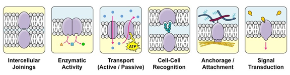
Figure 17: Membrane Protein Functions
I.5.3. Carbohydrates
Carbohydrates are covalently linked to proteins (glycoproteins) or lipids (glycolipids) and also an important part of cell membranes, and function as adhesion and address loci for cells.Membrane carbohydrates are chemically bound to glycolipids and glycoproteins.However, some membrane carbohydrates are part of proteoglycans that insert their amino acid chain among the lipid fatty acids.
Carbohydrate groups are present only on the outer surface of the plasma membrane and are attached to proteins, forming glycoproteins, or lipids, forming glycolipids.
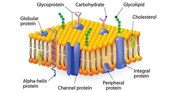
Figure 18 : Membrane Carbohydrate Structure
A-Glycoproteins
Glycoproteins are membrane proteins that have carbohydrate chains attached to them. Most membrane carbohydrates are found as part of glycoproteins, and nearly all membrane proteins contain sugars. These carbohydrates are usually short, branched oligosaccharides that provide specific recognition sites and enable high-affinity interactions with other molecules.
B-Glycolipids
Glycolipids are membrane lipids in which the hydrophilic head groups are oligosaccharides.Three types of glycolipids are found in membranes: glycosphingolipids, which are the most abundant in the animal cells, glycoglycerolipids, and glycophosphatidylinositol. Glycoglycerolipids are more frequent in the plasma membrane of plant cells. Only 5 % of lipids in membranes are glycolipids.
C- Proteoglycans
Polysaccharide chains of an integral membrane are called as proteoglycan molecules. Proteoglycans, which consist of long polysaccharide chains linked covalently to a protein core, are found mainly outside the cell as part of the extracellular matrix. But for some proteoglycans, the protein core either extends across the lipid bilayer or is attached to the bilayer by a glycosylphosphotidylinositol (GPI) anchor.
D-Glycocalyx
The glycocalyx (pl.: glycocalyces or glycocalyxes), also known as the pericellular matrix and cell coat, is an external organelle consisting of a layer of glycosylated biomolecules called glycoconjugates, such as glycoproteins and glycolipids.
These are embedded in and extend outwards from the cell membranes of virtually all cells.[1][2][3] Generally, the carbohydrate portion of the glycolipids found on the surface of plasma membranes helps these molecules contribute to cell–cell recognition, communication, and intercellular adhesion.
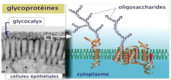
Figure 19 : Glycocalyx
I.6.Factors that influence membrane fluidity
Membrane fluidity is an essential property of biological membranes that allows proper membrane function and cellular activity. It depends on several physical and chemical factors related to the lipid and protein composition of the membrane.
1. Fatty acid chain length
Shorter fatty acid chains increase membrane fluidity, while longer chains
decrease it.
2. Degree of unsaturation
Unsaturated fatty acids (with double bonds) increase fluidity by preventing
tight packing of lipid tails. Saturated fatty acids decrease fluidity.
3. Cholesterol content
Cholesterol regulates membrane fluidity: it decreases fluidity at high
temperatures and increases fluidity at low temperatures by preventing lipid
packing.
4. Temperature
Higher temperatures increase membrane fluidity, whereas lower temperatures
reduce it.
5. Lipid composition
The types and proportions of phospholipids, glycolipids, and sphingolipids
affect how tightly the membrane components pack together.
6. Protein
content
The amount and type of membrane proteins can influence local membrane fluidity.
7. Presence
of lipid rafts
Lipid rafts, rich in cholesterol and sphingolipids, are more ordered and less
fluid than surrounding membrane regions.
I.7. Functions of Plasma Membrane
· The plasma membrane functions as a physical barrier between the external environment and the inner cell organelles.
· The plasma membrane is a selectively permeable membrane, which permits the movement of only certain molecules both in and out of the cell.
· The plasma membranes play an important role in both the endocytosis and exocytosis processes.
· The plasma membrane also functions by facilitating communication and signalling between the cells.
· The plasma membrane plays a vital role in anchoring the cytoskeleton to provide shape to the cell and also maintain the cell potential.