
Chapter 2: Substance Transport (Exchange mechanisms )
II.5.Active Transport
By the end of this section, you will be able to:Understand how electrochemical gradients affect ionsDistinguish between primary active transport and secondary active transport
Active transport mechanisms require the use of the cell’s energy, usually in the form of adenosine triphosphate (ATP). If a substance must move into the cell against its concentration gradient—that is, if the concentration of the substance inside the cell is greater than its concentration in the extracellular fluid (and vice versa)—the cell must use energy to move the substance. Some active transport mechanisms move small-molecular weight materials, such as ions, through the membrane. Other mechanisms transport much larger molecules.
II.5.1.Electrochemical Gradient
We have discussed simple concentration gradients—different concentrations of a substance across a space or a membrane—but in living systems, gradients are more complex. Because ions move into and out of cells and because cells contain proteins that do not move across the membrane and are mostly negatively charged, there is also an electrical gradient, a difference of charge, across the plasma membrane.
The interior of living cells is electrically negative with respect to the extracellular fluid surrounding them. At the same time, cells have a lower concentration of (Na+) than does the extracellular fluid. Therefore, both the concentration
gradient and the electrical gradient tend to drive Na+ into the cell. Conversely, cells have a higher concentration of K+ than the extracellular fluid does. Therefore, the concentration gradient tends to drive K+ out of the cell, while the electrical gradient tends to drive it inside the cell. The combined gradient of concentration and electrical charge that affects an ion is called its electrochemical gradient
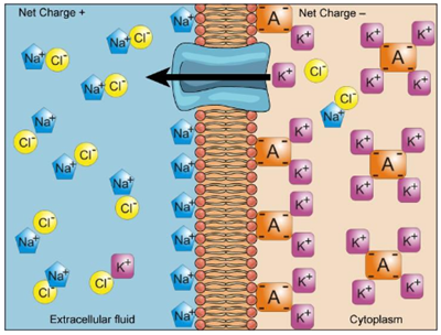
Figure 27: Electrochemical gradients arise from the combined effects of concentration gradients and electrical gradients.
II.5.2.Proteins for Active Transport
The specific proteins that facilitate active transport are called transporters. There are three types of transporters.
Ø A uniporter carries one specific ion or molecule.
Ø A symporter carries two different ions or molecules, both in the same direction.
Ø An antiporter carries two different ions or molecules in different directions. All of these transporters can transport small, uncharged organic molecules such as glucose.
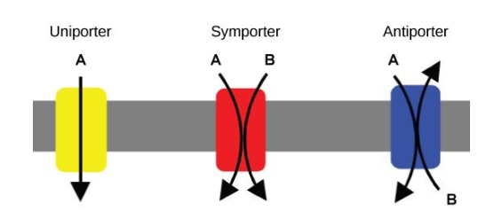
Figure 28 :A uniporter carries one molecule or ion. A symporter carries two different molecules or ions, both in the same direction. An antiporter also carries two different molecules or ions, but in different directions.
II.5.3.Primary Active Transport
One of the most important pumps in animals cells is the sodium-potassium pump (Na+-K+ ATPase), which maintains the electrochemical gradient and the correct concentrations of Na+ and K+ in living cells. The sodium-potassium pump moves two K+ into the cell while moving three Na+ out of the cell.
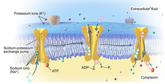
Figure 29 : Primary active transport moves ions across a membrane, creating an electrochemical gradient (electrogenic transport).
The sodium-potassium pump works in the following six steps:
1. Three sodium ions bind to the protein.
2. ATP is hydrolyzed by the protein carrier and a low-energy phosphate group attaches to it.
3. The carrier changes shape and opens towards the exterior of the membrane. The three sodium ions are released.
4. Two potassium ions attach to the protein, causing the low-energy phosphate group to detach.
5. The carrier protein changes shape so that is open towards the interior of the cell.
6. The two potassium ions are released into the cytoplasm and the process begins again.
II.5.4.Secondary Active Transport (Co-transport)
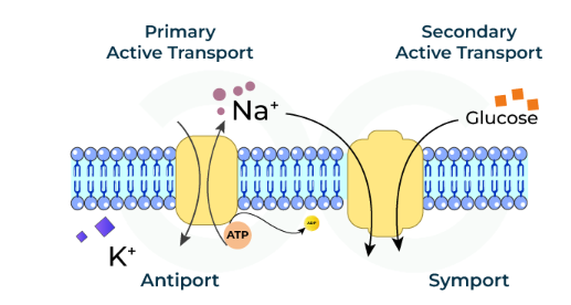
Secondary active transport moves a solute against its concentration gradient, an endergonic process, by moving another solute down its concentration gradient, an exergonic process. For instance, as sodium ion concentrations build outside of the plasma membrane because of the action of the sodium-potassium pump, an electrochemical gradient is created.
If a channel protein exists and is open, the sodium ions will be pulled through the membrane, down their concentration gradient. This exergonic movement is used to transport other substances that can attach themselves to the transport protein through the membrane . Many amino acids, as well as glucose, enter a cell this way.
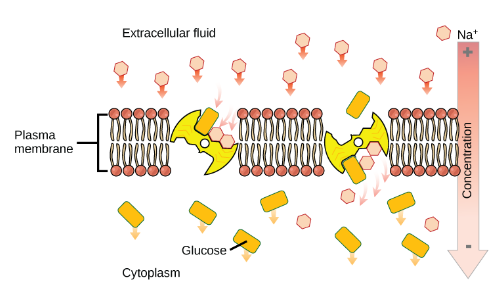
Figure 30: An electrochemical gradient, created by primary active transport, can move other substances against their concentration gradients, a process called co-transport or secondary active transport.
II.6.Bulk Transport
In addition to moving small ions and molecules through the membrane, cells also need to remove and take in larger molecules and particles. Some cells are even capable of engulfing entire unicellular microorganisms. You might have correctly hypothesized that the uptake and release of large particles by the cell requires energy. A large particle, however, cannot pass through the membrane, even with energy supplied by the cell.
II.6.1.Endocytosis
Endocytosis is a type of active transport that moves particles, such as large molecules, parts of cells, and even whole cells, into a cell. There are different variations of endocytosis, but all share a common characteristic: The plasma membrane of the cell invaginates, forming a pocket around the target particle. The pocket pinches off, resulting in the particle being contained in a newly created intracellular vesicle formed from the plasma membrane. The three types of endocytosis are phagocytosis, pinocytosis, and receptor-mediated endocytosis.
II.6.2.Phagocytosis
Phagocytosis (“cell eating”) is the process by which large particles, such as other cells or relatively large particles, are taken in by a cell. For example, when microorganisms invade the human body, a type of white blood cell called a neutrophil will “eat” the invaders through phagocytosis, surrounding and engulfing the microorganism, which is then destroyed by lysosomes inside the neutrophil .
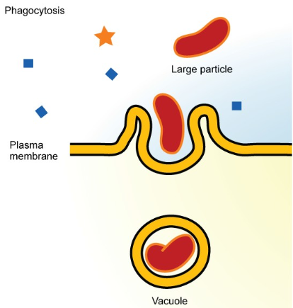
Figure 31: In phagocytosis, the cell membrane surrounds the particle and engulfs it.
II.6.3.Pinocytosis
Through pinocytosis (“cell drinking”), cells take in molecules, including water, which the cell needs from the extracellular fluid. Pinocytosis results in a much smaller vesicle than does phagocytosis, and the vesicle does not need to merge with a lysosome.
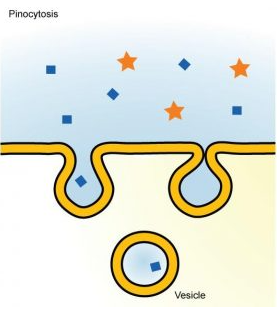
Figure 32 :In pinocytosis, the cell membrane invaginates, surrounds a small volume of fluid, and pinches off. (Credit: Mariana Ruiz Villareal)
Receptor-mediated Endocytosis
Receptor-mediated endocytosis is a targeted variation of endocytosis that employs receptor proteins in the plasma membrane that have a specific binding affinity for certain substances.
Receptor-mediated endocytosis, as in phagocytosis, uses clathrin protein attached to the cytoplasmic side of the plasma membrane. Some human diseases are caused by the failure of receptor-mediated endocytosis. For example, the form of cholesterol termed low-density lipoprotein or LDL (also referred to as “bad” cholesterol) is removed from the blood by receptor-mediated endocytosis. In the human genetic disease familial hypercholesterolemia, the LDL receptors are defective or missing entirely. People with this condition have life-threatening levels of cholesterol in their blood, because their cells cannot clear LDL particles from their blood.
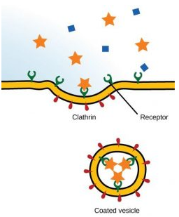
Figure 33 : In receptor-mediated endocytosis, uptake of substances by the cell is targeted to a single type of substance that binds to the receptor on the external surface of the cell membrane.
II.6.4.Exocytosis
The reverse process of moving material into a cell is the process of exocytosis. The purpose of exocytosis is to expel material from the cell into the extracellular fluid. Waste material is enveloped in vesicle, which fuses with the interior of the plasma membrane, expelling the waste material into the extracellular space. Cells also use exocytosis to secrete proteins such as hormones, neurotransmitters, or parts of the extracellular matrix.
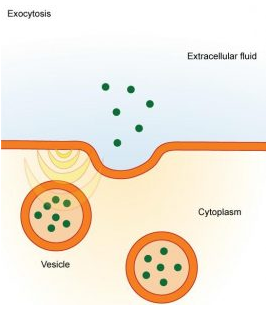
Figure 34 : In exocytosis, vesicles containing substances fuse with the plasma membrane. The contents are then released to the exterior of the cell.
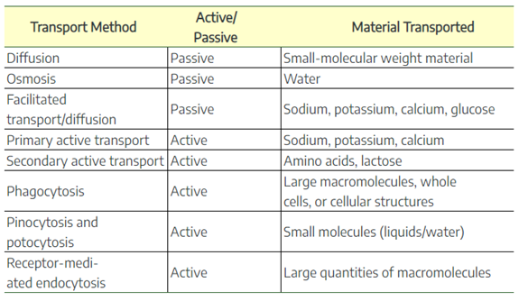
Table 01 : Methods of transport, energy requirements, and types of material transported
II.7.Factors Influencing Transport Across Cell Membrane
There are four factors that influence the process of transportation, and these factors include:
1. Gradient concentration: Transportation of molecules usually occurs from a high concentration gradient to a low concentration, but when transportation occurs from a low concentration to a high concentration, it requires energy.
2. Size of the molecules: Molecule size also affects the rate of transport; the smaller the size, the faster the movement.
3. Lipid Soluble Molecules: Molecules that are lipid-soluble can be transported through a lipid bilayer.
4. Charge on molecule: Molecules that have charge on them require carrier proteins for transportation.
II.8.Examples of Transport Across the Cell Membrane
Here are real-life examples of different kinds of transport happening in the body:
1. Simple diffusion: Exchange of carbon dioxide and oxygen across the lipid bilayer.
2. Facilitated diffusion: Transportation of glucose via a glucose transporter.
3. Osmosis:The movement of water across the cell membrane.
4. Primary active transport: Transportation of Na+ and K+ ions from inside to outside of the cell and from outside to inside of the cell, respectively.
5. Secondary active transport: Transportation of glucose against a concentration gradient via a sodium-glucose co-transporter.