Chapter I : The membrane ultrastructure (part 1)
I.1.IntroductionThe plasma membrane, which is also called the cell membrane, has many functions, but the most basic one is to define the borders of the cell and keep the cell functional. The plasma membrane is selectively permeable. This means that the membrane allows some materials to freely enter or leave the cell, while other materials cannot move freely, but require the use of a specialized structure, and occasionally, even energy investment for crossing.
I.2.Plasma membrane
The cell membrane (also known as the plasma membrane (PM) or cytoplasmic membrane, and historically referred to as the plasmalemma) is a biological membrane that separates the interior of all cells from the outside environment (the extracellular space) which protects the cell from its environment.
The model was described as a ‘lipo-protein sandwich’, as the lipid layer was sandwiched between two protein layers.The dark segments seen under electron microscope were identified (wrongly) as representing the two protein layers.When viewed under a transmission electron microscope, membranes exhibit a characteristic ‘trilaminar’ appearance
Trilaminar
= 3 layers (two dark outer layers and a lighter inner region)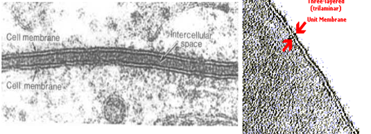
Figure 01: Low- and high-magnification transmission electron micrographs of the trilamellar membrane.
I.3.Fluid Mosaic Model
The existence of the plasma membrane was identified in the 1890s, and its chemical components were identified in 1915. The principal components identified at that time were lipids and proteins. The first widely accepted model of the plasma membrane’s structure was proposed in 1935 by Hugh Davson and James Danielli; it was based on the “railroad track” appearance of the plasma membrane in early electron micrographs.
They theorized that the structure of the plasma membrane resembles a sandwich, with protein being analogous to the bread, and lipids being analogous to the filling. In the 1950s, advances in microscopy, notably transmission electron microscopy (TEM), allowed researchers to see that the core of the plasma membrane consisted of a double, rather than a single, layer.
A new model that better explains both the microscopic observations and the function of that plasma membrane was proposed by S.J. Singer and Garth L. Nicolson in 1972.
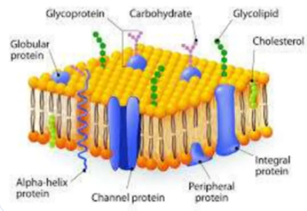
Figure 02: fluid mosaic model.
The explanation proposed by Singer and Nicolson is called the fluid mosaic model. The model has evolved somewhat over time, but it still best accounts for the structure and functions of the plasma membrane as we now understand them. The fluid mosaic model describes the structure of the plasma membrane as a mosaic of components—including phospholipids, cholesterol, proteins, and carbohydrates—that gives the membrane a fluid character. Plasma membranes range from 5 to 10 nm in thickness. For comparison, human red blood cells, visible via light microscopy, are approximately 8 µm wide, or approximately 1,000 times wider than a plasma membrane. The membrane does look a bit like a sandwich.
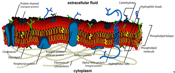
Figure 03 : This model of the eukaryotic plasma membrane demonstrates membrane asymmetry, with a sugarless cytoplasmic surface and a glycoprotein-rich extracellular surface.
I.4.Caractrestic of The fluid mosaic model
The fluid mosaic model describes how the molecules are arranged within cell membranes
Ø The term "fluid" refers to the lateral movement of phospholipids and some proteins, giving the membrane flexibility
Ø The term "mosaic" reflects the scattered arrangement of proteins within the bilayer
This model explains how membranes are:
Ø Partially permeable
Ø Sites for cell signalling, recognition, and communication
Ø Responsible for controlling the exchange of substances across compartments
The basic structure of all cell membranes is the same. This includes the cell surface membrane and the membranes surrounding eukaryotic organelles (e.g. nucleus, mitochondria, endoplasmic reticulum, Golgi apparatus, chloroplasts, lysosomes)
These membranes:
Ø Are composed of a phospholipid bilayer
Ø Contain intrinsic and extrinsic proteins
Ø May include cholesterol (in animal cells), glycoproteins, and glycolipids.
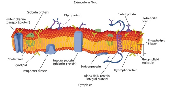
Figure04 : Schematic Diagram of
a Cell Membrane.
The membrane enclosing a typical animal cell is a phospholipid bilayer with embedded cholesterol and protein molecules. Short oligosaccharide chains are attached to the outer surface.
I.5.Chemical structure of plasma membrane
I.5.1.Membrane lipids
lipid constitutes 40% of the mass of most cell membranes, although this proportion varies depending on the type of cell. Include phospholipids and cholesterol.
I.5.1.1.Phospholipids
The fundamental building blocks of all cell membranes, which are amphipathic molecules , consisting of two hydrophobic fatty acid chains linked to a phosphate- containing hydrophilic head group.
A. The hydrophilic (polar) heads of the phospholipids molecules face the intracellular and extracellular fluids, consisting of:
· Phosphoric acid group
· Glycerol backbone
B. The hydrophobic (non polar) tail face each other in the membrane interior, consist of:
· Saturated fatty acid
· Unsaturated fatty acid, this double bond introduces a kink in the chain which reduces phospholipid packing (double bond increase fluidity).
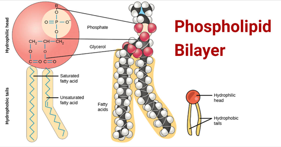
Figure 05: Phospholipid bilayer depicting hydrophobic tails of fatty acid chains and hydrophilic head of glycerol backbone, phosphate, and polar head group.
Three classes of complex amphiphilic lipids make up biological membranes:
· Glycerophospholipids
· Sphingolipids
· Steroids
· Glycolipids
I.5.1.2.Glycerophospholipids
They represent the first major class of lipids in the bilayer of the plasma membrane. Glycerophospholipids have a hydrophilic polar head composed of an alcohol R′ (choline, ethanolamine, inositol, serine, etc.), a phosphate group, and glycerol (an alcohol), and a hydrophobic, nonpolar tail made up of two hydrocarbon fatty acid chains.
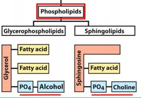
Figure 06 : glycerophospholipid
Different classes of glycerophospholipids
Eukaryotic phospholipids are of several types. Four major glycerophospholipids are mainly found in membranes:
1. Phosphatidylserines (9%)
2. Phosphatidylethanolamines (11%)
3. Phosphatidylcholines (lecithin) (18%)
4. Phosphatidylinositols (low abundance)
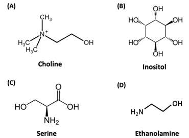
Figure07 :. A few of the common components of phosopholipid head groups. (A) Choline, (B) inositol, (C) serine, and (D) ethanolamine are all hydrophilic groups that can be attached to the phosphate in phospholipids.
I.5.1.3.Sphingolipids
Sphingolipids are a type of phospholipid that have a hydrophobic tail made up of only one fatty acid chain and a sphingosine molecule. Sphingosine is derived from the amino acid serine and a long hydrocarbon chain; this hydrocarbon chain runs parallel to the fatty acid chain in a sphingolipid. Like phospholipids, the hydrophilic head group on sphingolipids can vary. An example of the chemical structure of a sphingolipid can be viewed in the carbohydrates section below when discussing glycolipids.
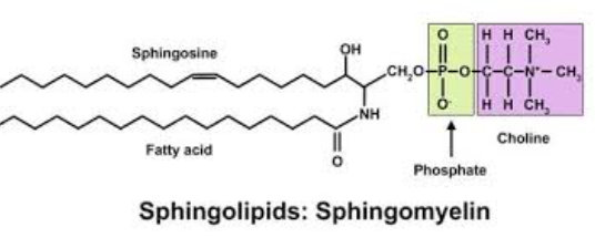
Figure08 : sphingolipids
I.5.1.4. Sterols
Sterols are another type of lipid found in the cell membrane. They are multi-ringed structures that are mostly hydrophobic, except for a hydrophilic hydroxyl group. In the membrane their hydrophobic regions associate with phospholipid tails, and their hydrophilic regions associate with phospholipid head groups.
I.5.1.5.Cholesterol
Cholesterol belongs to a category of lipids called steroids. It is an essential component of all animal cell membranes (the lipid bilayer). Within these membranes, cholesterol helps to maintain the membrane’s structure so that cells can function effectively. We will focus on the structural role of cholesterol and its impact on membrane fluidity as demonstrated through experimentation. However, it is also important to note that cholesterol plays other significant roles, such as the biosynthesis of steroid hormones like testosterone and estrogen.
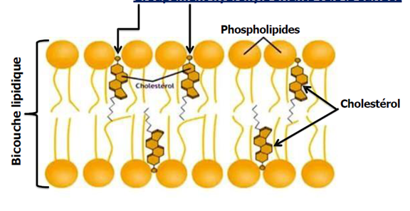
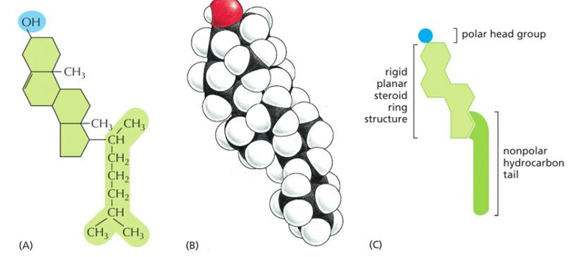
Figure 09 : cholesterol
Cholesterol’s structure
Similar to phospholipids in the cell membrane, cholesterol displays amphipathic properties. In other words, it possesses a polar head that contains a hydrophilic hydroxyl group, while the remainder of the molecule is hydrophobic, consisting of a 4 fused carbon ring structure and a nonpolar hydrocarbon tail